

Large-scale analysis of lipoprotein(a) levels in a UK-wide population, including analyte stability and comparability between capillary and venous samples: A feasibility study for home self-testing for personalized cardiovascular risk management
Abstract
Background & aim: Lipoprotein(a) (Lp(a)) is an established predictor of cardiovascular risk. Recent evidence suggests that Lp(a) testing with subsequent intervention could be economically advantageous, and several organizations now recommend population testing. However, general population datasets are scarce, and healthcare services already under pressure may not be able to undertake large-scale testing programs using traditional sampling methods, even when financially beneficial. However, self-collected capillary blood samples offer several advantages over venous collections, primarily by enabling patients to collect samples whenever and wherever they choose, removing the phlebotomist bottleneck. This patient-centric approach has become increasingly popular because of its convenience. Evidence regarding the comparability of venous and capillary samples for various routine analytes remains limited. Moreover, questions remain about the stability of these analytes in whole-blood samples over time, particularly when separation is delayed before analysis. This project aimed first to review a UK-wide Lp(a) dataset and compare it with other recently published UK Biobank data, and second to determine whether screening could be performed using capillary blood. This was done by investigating venous and capillary comparability and sample stability in various anticoagulants over 72 hours. Materials & methods: Serum Lp(a) concentrations from 5298 venous samples collected between January 2025 and June 2025 were provided from a UK-wide population dataset and compared with a recently published UK Biobank dataset. Stability studies were organized as follows. Five paired venous and capillary serum samples were collected and tested on day zero to serve as an initial baseline verification. We collected a further 22 paired capillary and venous blood samples. The venous sample was tested on day zero, and the capillary sample was stored as whole blood at room temperature and processed 72 h later. A further 10 samples were obtained to confirm day-zero and 72 h comparability between venous serum and EDTA plasma samples. Results were compared using Pearson’s correlation, Bland–Altman and Passing–Bablok analyses. Results: Statistical analysis showed that Lipoprotein (a) levels in capillary and venous serum and plasma samples were comparable, and that the analyte was stable in unspun blood across all the matrices tested for at least 72 h before analysis. Conclusion: We believe this is the first published study to determine the comparability and stability of venous and capillary measurements of Lp(a). This study indicates that Lp(a) analysis is feasible using self-collected capillary samples returned to the laboratory via the postal system.
Plain language summary
Lipoprotein(a), or Lp(a), is a type of fat-and-protein particle made by the liver that carries cholesterol through the bloodstream; the amount people have is largely determined by genetics. High levels are associated with an increased risk of cardiovascular disease. Wider testing could help identify people at increased risk, but population-scale screening using traditional blood collection methods, venepuncture, may place additional pressure on healthcare services.
This study examined whether Lp(a) could be measured using capillary blood samples that people may collect themselves at home. Lp(a) results from 5298 venous blood samples collected across the UK were reviewed and compared with published UK Biobank data. The study also compared Lp(a) measurements from paired capillary and venous samples and assessed whether Lp(a) remained stable when whole blood was stored at room temperature for up to 72 h before processing.
Lp(a) measurements were comparable across capillary and venous serum and plasma samples. Lp(a) also remained stable in unprocessed blood for at least 72 h in all the sample types tested.
These findings suggest that self-collected capillary blood samples could be returned to a laboratory by post and used reliably for Lp(a) testing. This approach could make wider Lp(a) screening more convenient and accessible, while reducing reliance on traditional phlebotomy services.
Cardiovascular diseases (CVD) are the leading cause of death worldwide [1–3]. The economic and healthcare burden of CVD is estimated at US $400 billion in the US and over €280 billion in the EU annually [1,4].
The worldwide incidence and cost of CVD are increasing rapidly, driven by an ageing global population and limited availability of primary and secondary prevention programs [5]. It is therefore essential that nations prioritize CVD prevention and management strategies [6].
Well-established cardiovascular risk factors include high LDL cholesterol, high blood pressure, diabetes, obesity, smoking, physical inactivity and an unhealthy diet [7,8]. In addition, low socioeconomic status and gender are strongly associated with CVD risk [9,10]. More recently, elevated lipoprotein (a) (Lp[a]) has also been recognized as an independent risk factor for CVD [11].
Lp(a) consists of an LDL-like particle with an added apolipoprotein and contains a significant number of oxidized phospholipids, which are thought to contribute to its atherogenic and inflammatory effects. Lp(a) levels are primarily determined by genetics, with approximately 90% of the variance explained by an individual’s genetic makeup. This makes a high Lp(a) level the most commonly inherited dyslipidaemia in humans [11–14].
The Lp(a) Brussels International Declaration Summit identified an urgent need for systematic Lp(a) testing as part of personalized cardiovascular risk management [14]. Although the cost savings of such testing regimes are difficult to assess, a recent paper found that, from a societal perspective, Lp(a) testing would save the UK £263 per person [15].
Many Lp(a)-lowering drugs, including monoclonal antibodies and antisense oligonucleotides, are in clinical trials and show promise [16]. Increased Lp(a) testing could therefore identify high-risk individuals, enable targeted treatment and prevent many cardiovascular events, thereby saving substantial downstream healthcare costs [17].
There is a notable size heterogeneity that hampers accurate quantification of the mass of the Lp(a) molecule; consequently, the WHO/IFCC recommendations are that assays be reported in molar units (nmol/l) [18,19]. To date, not all manufacturers have updated their assays in line with this guidance.
In a large UK Biobank dataset, Kao et al. found that Lp(a) is independently associated with ischemic stroke, with variation by sex but not by age or race/ethnicity. The prevalence of Lp(a) >125 nmol/l was 11.1%, and this level is considered to confer a high risk of future CVD [20]. In addition, McClintick et al. recently found that among 3757 individuals with peripheral artery disease, those with Lp(a) levels of 42–<132 nmol/l and 132–855 nmol/l had a 24% and 30% increased hazard of a major adverse cardiovascular event, respectively, compared with those with levels below 42 nmol/l [21].
Although lipid status is typically assessed using venous serum, conventional venipuncture is not always feasible or preferred by the patient, and its widespread use for large-scale population screening would be financially prohibitive. As such, alternative sampling strategies, including capillary sampling, could support a more patient-centric, cost-efficient and effective CVD prevention and management strategy. Recently published evidence by Hoffmeister et al. suggests that routine lipid analytes, total cholesterol, triglycerides, HDL and LDL, alongside HbA1C, were both stable and comparable in capillary blood [22]. Because these analytes are commonly used to evaluate CVD risk [23], establishing whether Lp(a) could be included in self-collected capillary blood testing programs may be of interest. This is especially true if the analysis could be performed using either serum as part of the lipid profile panel or using plasma from the HbA1C sample.
Capillary sampling has several advantages over venous sampling, including being minimally invasive, requiring fewer raw materials, and reducing waste in both plastics and blood. It also enables large numbers of people to be tested efficiently and cost-effectively, as neither a clinical site nor staff are required for sample collection.
A growing body of published work supports capillary blood testing. Published data include comparisons of the complete blood count, enzymatic creatinine, urea, liver function tests, lipids, CRP, HbA1C, vitamin D, PSA, AMH, LH, FSH, estradiol, testosterone, prolactin, DHEA-S, SHBG, TSH and FT4 [24–31]. As such, this sample-collection method is gaining increasing interest among healthcare providers [32].
This proof-of-concept study first reviewed Lp(a) results from the laboratory for the previous 6-month period. This was done to determine whether patients self-referring for profiles that include Lp(a) were representative of those reported elsewhere [20]. These data come from a UK-wide wellness population, i.e., self-referring individuals. We then investigated whether self-collected capillary blood methods, with return to the laboratory for Lp(a) testing, were feasible.
This second aim was to determine whether venous and capillary Lp(a) concentrations were comparable on day 0 and at 72 h. Stored samples were kept unseparated at 20–24°C. We chose the 72 h period to mimic postal timing; we also inverted each whole-blood sample five times per day to mimic transport conditions. Finally, paired serum and plasma venous samples were selected to compare whole-blood plasma Lp(a) concentrations over 72 h with serum Lp(a) concentrations. The serum sample was analyzed on day 0, and the whole-blood sample was stored at 20–24°C for 72 h before processing.
Materials & methods
Sample collection & preparation
All participants received diagrammatic guidance on performing effective capillary sampling, and self-collection was performed immediately after venous sampling. A trained phlebotomist collected venous samples using Becton Dickinson (Oxford, UK) Serum Separator Tubes (SST), while capillary samples were collected using Becton Dickinson Microtainer SST tubes and 2 mm self-retracting safety lancets. As part of the project, one pair of matched venous and capillary samples was centrifuged together at 2000 g for 10 min within 4 h of collection and analyzed immediately on the Roche Cobas c503 (Roche Diagnostics, Burgess Hill, UK). The second set of whole-blood capillary samples was stored at room temperature (20–24°C) for 72 h before centrifugation and then tested together on the same platform. We inverted the samples five times each day to resuspend the cells and mimic transport conditions. Lp(a) was analyzed according to the manufacturer’s recommendations. Samples were excluded based on the hemolytic, icterus and lipemia indices as reported by the analyzer (Roche Diagnostics).
Twenty-two generally well patients (17 females and 5 males, aged 21–52 years) who had requested Lp(a) testing as part of a wellness panel were asked to provide one venous and one or two capillary samples. We also used an additional ten participant samples to assess serum and plasma comparability in venous blood. The number of participants aligns with the lower limit of comparison samples specified by both the Clinical and Laboratory Standards Institute document on the assessment of equivalence of specimen types for medical laboratories [33] and recent UK recommendations for validating capillary blood samples [34], which state that a minimum of 20 paired samples is required with justification. A final group of ten participants gave two venous samples, one serum and one EDTA. The serum sample was tested on day 0, and the EDTA sample was stored for 72 h with the whole-blood capillary samples and analyzed with them. All aspects of this study were conducted in accordance with the Declaration of Helsinki (2013), and individuals were included only after informed consent.
Roche Tina-quant assay (Gen 2)
The Roche Tina Quant Lp(a) test is a 2-end-point particle-enhanced immunoturbidimetric assay, where human Lipoprotein (a) agglutinates with latex particles coated with anti-Lp (a) antibodies. The precipitate is determined turbidimetrically at 800 and 660 nm. The standard limit of detection for this assay is 7–240 nmol/l. Samples with higher concentrations are diluted 1:3, resulting in a higher limit of detection of 720 nmol/l.
However, to fully utilize this technology for population screening and CVD management, comparability between venous and capillary samples, as well as whole-sample stability, is critical. To date, there is limited data for either of these two variables for Lp(a). The current Roche IFU states that venous samples should be assayed within 8 h; if this is not possible, samples should be stored at 2–8°C. If samples are not assayed within 48 h, they should be stored frozen at -70°C (±10°C) or below. Wigh et al. recently published evidence supporting venous whole blood stability for Lp(a) for up to 10 h at 21°C [30]. Here, we provide additional evidence for the stability of Lp(a) in whole-blood capillary samples stored between 20 and 24°C for up to 72 h. We have also provided additional evidence using whole-blood plasma samples rather than serum from SST.
Statistical analysis
Pearson’s Correlation, Bland–Altman and Passing–Bablok analyses were used to assess the strength of the relationship and the level of agreement between Lp(a) concentrations in venous blood from samples taken on day 0 and capillary samples tested at 72 h, as described by Bland et al. and Thienpont et al. [35,36]. All statistical analyses were performed using Analyse-It Method Validation Edition software (Analyse-It, Leeds, UK). Acceptance criteria were a correlation score of r >0.95 and a Bland–Altman result within the total allowable error for Lp(a). The analytical within-subject coefficient of variation of Lp(a) is 10% (95% CI: 4.2%–26.7%), and the total allowable error is quoted as being 15% [37].
Results
The laboratory’s information system was searched for all patient Lp(a) results from the prior 6 months. The total number of patient records, including Lp(a) results, was 5298; 2100 (approx. 40%) were female, and 3198 (approx. 60%) were male (Tables 1 & 2).
| Female n = 2100 | |
|---|---|
| Age range (years), 18–94 | |
| Lp(a) range <7–664 nmol/l | |
| Lp(a) nmol/l | Patients (n) |
| <25 | 1152 |
| 25–50 | 286 |
| 51–75 | 118 |
| 76–125 | 172 |
| 126–150 | 64 |
| 151–175 | 65 |
| 176–200 | 67 |
| 201–250 | 109 |
| 251–300 | 25 |
| 301–350 | 18 |
| 351–400 | 12 |
| 401–500 | 6 |
| >500 | 6 |
| Male n = 3198 | |
|---|---|
| Age range (years), 18–85 | |
| Lp(a) range <7–612 nmol/l | |
| Lp(a) nmol/l | Patients (n) |
| <25 | 1743 |
| 25–50 | 416 |
| 51–75 | 189 |
| 76–125 | 250 |
| 126–150 | 138 |
| 151–175 | 116 |
| 176–200 | 93 |
| 201–250 | 153 |
| 251–300 | 51 |
| 301–350 | 18 |
| 351–400 | 17 |
| 401–500 | 10 |
| >500 | 4 |
To determine whether capillary and venous Lp(a) concentrations were comparable and whether Lp(a) levels were stable in whole blood capillary samples during a typical postal time period, we collected paired venous and capillary samples from 22 study participants aged 21–52 years. As a baseline, we collected two capillary samples from five patients whose results spanned a wide clinical range; we tested one alongside the venous sample on day 0 to confirm same-day comparability (Table 3). We stored the remaining capillary samples unprocessed as whole blood for 72 h at 20–24°C before centrifugation and testing. We mixed stored samples by 5 inversions per day to fully resuspend the cells. This was done to mimic postal conditions.
| Venous day 0 | Capillary day 0 |
|---|---|
| <7 | <7 |
| 7 | 7 |
| 107 | 100 |
| 134 | 126 |
| 210 | 219 |
Lp(a) measurements in venous and capillary blood tested on day 0 (units nmol/l, n = 5). Correlation between venous and capillary serum from day 0 is r = 0.997
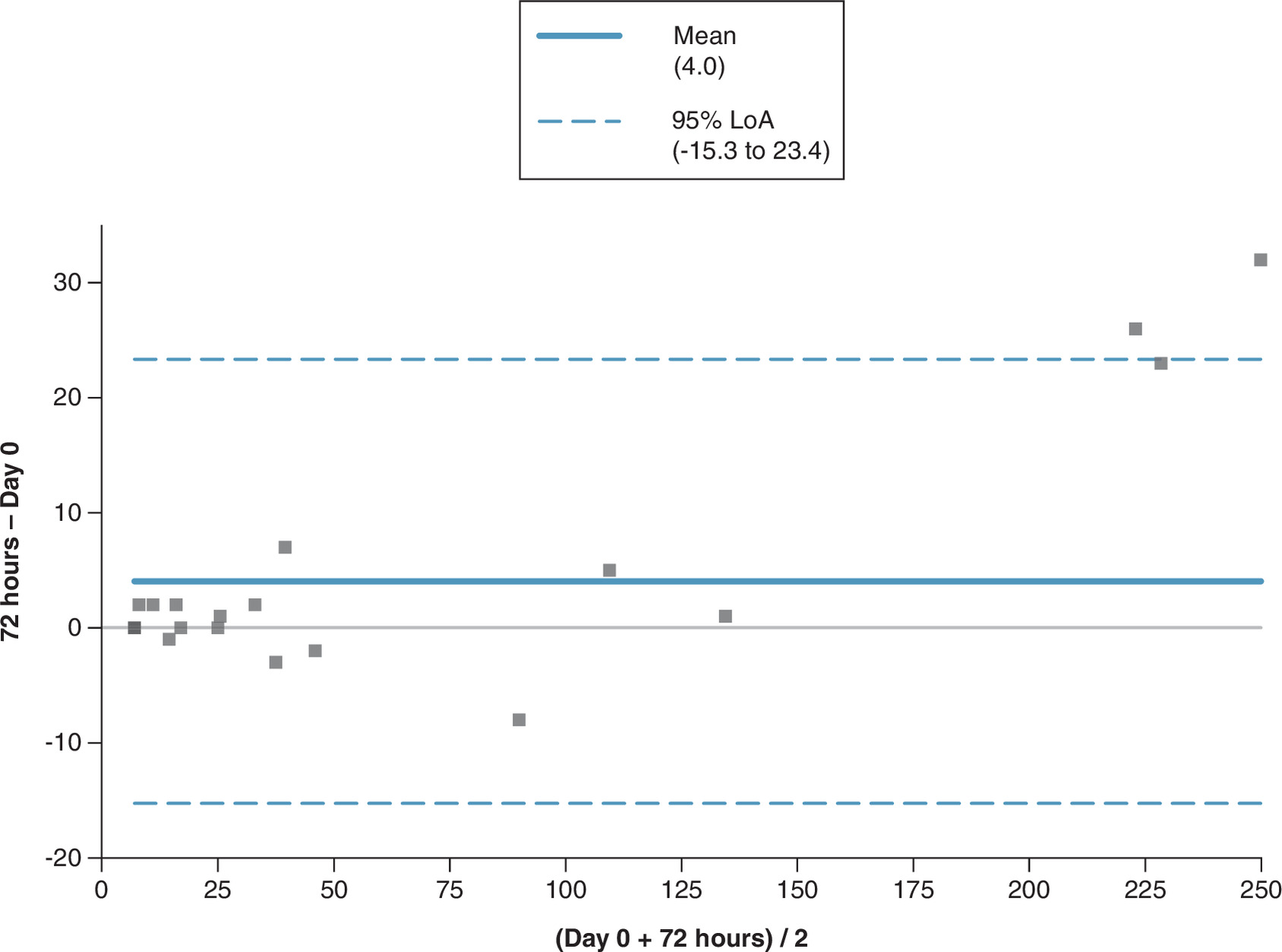
Table 3 shows that day 0 capillary and venous results were highly comparable and correlated (r = 0.997). An average bias of 4.0 nmol/l was observed between 72 h-old whole-blood capillary samples and day 0 venous serum samples (Table 4), within the optimal total allowable error of 15% [37]. A similar correlation was observed when day 0 serum samples were compared with whole blood EDTA samples stored for 72 h before processing (Table 5).
| Venous day 0 | Whole blood capillary 72 h |
|---|---|
| <7 | <7 |
| <7 | <7 |
| <7 | <7 |
| <7 | <7 |
| <7 | <7 |
| 7 | 9 |
| 10 | 12 |
| 15 | 14 |
| 15 | 17 |
| 17 | 17 |
| 25 | 26 |
| 25 | 25 |
| 32 | 34 |
| 36 | 43 |
| 39 | 36 |
| 47 | 45 |
| 94 | 86 |
| 107 | 112 |
| 134 | 135 |
| 210 | 236 |
| 217 | 240 |
| 234 | 266 |
Lp(a) measurements in venous serum from day zero and capillary samples tested at 72 h after collection (units nmol/l, n = 22). Pearson’s correlation between venous day 0 and capillary at 72 h, r = 0.998.
| Venous SST day 0 | Venous EDTA 72 h |
|---|---|
| <7 | <7 |
| <7 | 7 |
| 21 | 22 |
| 23 | 24 |
| 25 | 25 |
| 74 | 77 |
| 85 | 88 |
| 160 | 166 |
| 204 | 226 |
| 386 | 395 |
Lp(a) measurements in venous serum from day 0 and venous EDTA samples tested at 72 h after collection (units nmol/l, n = 10). Correlation: r = 0.999.
The Bland–Altman plot (Figure 1) comparing Lp(a) concentrations from venous samples tested on day 0 and capillary samples tested at 72 h indicates a positive mean bias of 4.0 nmol/l.
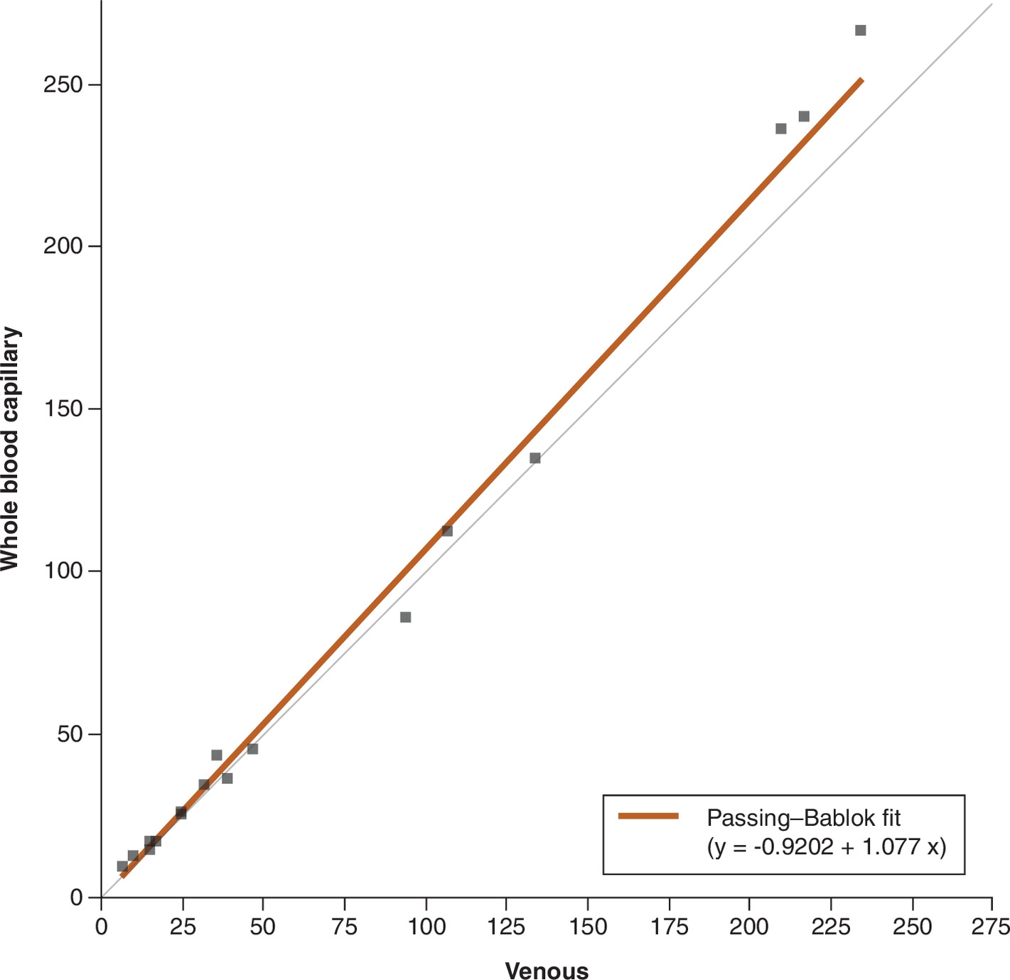
The Passing–Bablok plot of venous and capillary serum determinations of Lp(a) concentration on day zero (correlation coefficient r = 0.998, n = 22) shows a constant negative bias of 0.9 nmol/l and a 7.7% positive proportional bias. No significant difference between venous and capillary serum is identified, as the 95% CI encompasses 1 for the slope and 0 for the intercept.
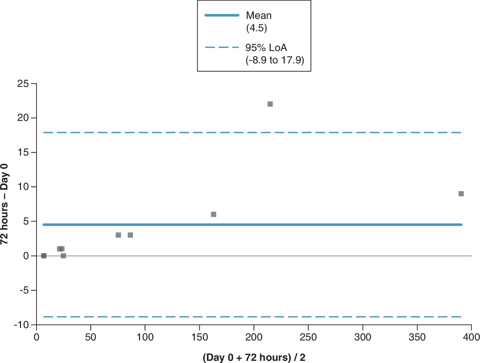
A Bland–Altman plot (Figure 2) comparing Lp(a) concentration from venous serum samples tested on day 0 and venous EDTA samples tested at 72 h indicates a positive mean bias of 4.5 nmol/l.
Discussion
Venepuncture remains the gold standard for blood sampling and is among the most frequently performed medical procedures in clinical practice. However, the need for specialist personnel and equipment limits its use in some circumstances and may contribute to bottlenecks in the patient pathway. Additionally, the environmental costs of phlebotomy, including patient travel, consumables, sample storage and disposal, may be substantial. NHS England found that up to 90% of the carbon footprint of laboratory tests is attributable to the sample collection process [38].
Self-collected capillary blood sampling with return-to-laboratory analysis has proven to be a viable alternative to venipuncture for a wide range of analytes [31] and could be a significant contributor to a more patient-centric, personalized, cost-efficient healthcare system [39], focused on patient participation and disease prevention; it could also significantly reduce the environmental impact of the blood collection process and blood wastage. However, its use will be limited without comparability and analyte stability studies [40].
Raised Lp(a) levels have been identified as a major independent risk factor in the pathophysiology of CVD, and early detection is considered a critical part of personalized cardiovascular risk management [14,41,42]. Accordingly, it is recommended that all adults undergo Lp(a) testing and that cascade screening be used to identify elevated Lp(a) levels in relatives [41,42]. Lp(a) testing would likely be performed alongside more traditional markers, such as lipids and HbA1C; however, traditional healthcare systems are unlikely to screen a large population using venous sampling pathways. The time and resource costs would be prohibitive, especially given the current strains on these services. The comparability of standard lipids and HbA1C between capillary and venous blood has largely been confirmed [22,25,29]. Because Lp(a) can be measured from either serum or plasma using self-collected capillary blood samples, barriers to large-population testing could be removed if comparability and stability in whole blood are confirmed.
The population data presented above in Tables 1 & 2 suggest that 18% of self-referring patients had Lp(a) concentrations >125 nmol/l, which is higher than the recently published UK Biobank study [20], which reported that approximately 11% of individuals had Lp(a) levels >125 nmol/l. Our data suggest that almost 20% of the patients tested had an Lp(a) level in the high-risk category, aligning with data published by Nissen et al., who studied patients with established ASCVD [42]. Our study included just over 5000 patients, which is significantly fewer than in Kao et al. [20]. Published long-term evidence supporting Lp(a) stability at -70°C is limited [43]; therefore, the data in Tables 1 & 2 may more accurately reflect true levels in the UK population.
The population included in Tables 1 & 2 is self-referring and may therefore be drawn from a higher socio-economic patient group. However, given that Lp(a) is largely under genetic control and that these samples are from a UK-wide population, the self-referring nature of the study population is unlikely to be significant, as these samples came from preventative healthcare blood profiles. However, self-referral due to a family history of early heart disease cannot be ruled out.
To ensure accurate results, it is essential to validate the comparability between venous and capillary blood and to determine analyte stability during routine transport times [34]. Currently, no published data compare venous and capillary blood for Lp(a) using standard laboratory assays, and whole blood stability data are limited to hours rather than days [30].
This feasibility study tested five paired venous and capillary serum samples, spanning low- to high-risk levels, to verify comparability on day 0. Samples spanned the assay's lower limit of detection (<7 nmol/l) to levels considered high risk (>125 nmol/l). We then analyzed a further 22 paired venous and capillary samples, with the venous sample tested on day 0 and the capillary samples stored unspun for 72 h at 20–24°C before analysis.
The results from capillary and venous samples were highly comparable and correlated (r = 0.998). The average bias between venous and stored capillary samples was 4.0 nmol/l (Figure 1), which is within the optimal total allowable error of 15%. Ten additional paired venous serum and EDTA samples confirmed that Lp(a) is also stable in this matrix for the same time period, with a 4.5 nmol/l positive bias (Figure 3).
These data also indicate that Lp(a) remains stable in serum and EDTA plasma for at least 72 h at room temperature (20–24°C), suggesting that self-collected capillary samples using either serum or EDTA tubes could be used to assess patients’ Lp(a) levels via standard postal services. Our data suggest that self-collected capillary samples could be not only highly effective at identifying high-risk individuals but also highly efficient.
This study is the first to present data showing that Lp(a) levels are comparable in venous and capillary samples across a broad physiological range and that they remain stable for up to 72 h at temperatures between 20 and 24°C.
These results are particularly important when investigating populations where there are concerns about using more invasive venous sampling techniques or the costs and resources required for phlebotomy. This sampling method reduces the need for patients to visit primary care or outpatient clinics for blood tests, potentially lowering nonattendance rates and supporting underserved populations.
However, the study has several limitations. We have controlled only for the transport time routinely used by the UK’s Royal Mail Tracked 24 service, in which over 95% of samples are returned within 72 h (own unpublished data). Likewise, because serial capillary samples are difficult to obtain, we could perform stability studies only within a limited temperature range. The data presented cover a wide range of Lp(a) concentrations (<7–266 nmol/l in serum and <7–395 nmol/l in plasma). Although based on a limited dataset, four of the 22 (18%) study participants had Lp(a) results over 125 nmol/l, the level considered to confer a high risk of future CVD [20], and comparable to the population data sets given above in Tables 1 & 2.
Conclusion
This study demonstrates that a general UK population has higher Lp(a) levels than previously reported by the UK BioBank [20], with concentrations consistent with those observed by Nissen et al. [42]. Furthermore, the findings indicate that Lp(a) concentrations are comparable between capillary and venous blood samples and that Lp(a) remains stable in whole-blood capillary samples for at least 72 h, supporting the potential use of capillary blood testing for self-collected samples in laboratory-based Lp(a) analysis.
Summary points
•
Lipoprotein(a) or Lp(a), is a genetically determined and independent cardiovascular risk factor, with elevated concentrations associated with an increased risk of cardiovascular disease.
•
The study analyzed 5298 Lp(a) results collected from a UK-wide, self-referred wellness-testing population between January and June 2025.
•
Approximately 18.3% of the study population had an Lp(a) concentration above 125 nmol/l, a threshold associated with high cardiovascular risk.
•
The proportion above 125 nmol/l was similar in women and men: approximately 17.7% and 18.8%, respectively.
•
The observed prevalence was higher than the approximately 11% reported in the referenced UK Biobank study, although differences in population selection and study methods limit direct comparison.
•
Same-day measurements from five paired venous and capillary serum samples showed close agreement across an Lp(a) range of below 7–219 nmol/l.
•
In 22 paired samples, capillary whole blood stored unprocessed at 20–24°C for 72 h showed strong correlation with venous serum tested on the day of collection.
•
The reported mean positive bias between day-zero venous serum and 72 h capillary samples was approximately 4 nmol/l, within the study’s predefined total allowable error.
•
Ten paired venous samples also showed close agreement between day-zero serum and EDTA plasma processed after 72 h, with a mean positive bias of approximately 4.5 nmol/l.
•
The findings support the feasibility of using self-collected capillary samples for laboratory-based Lp(a) measurement, but larger real-world studies involving postal transport, broader temperatures and more diverse populations are required.
Author contributions
T Woolley was responsible for the drafting and revision of the manuscript. E Rutter/L Fitzgerald/N Young contributed to the redrafting, statistical review and subsequent revisions. All authors reviewed and edited the manuscript and approved the final version.
Financial disclosure
The authors received no financial and/or material support for this research or the creation of this work.
Competing interests disclosure
Timothy Woolley, Emma Rutter and Leanne Fitzgerald are employed by Inuvi Health; Noel Young is employed by Thriva.co. The authors have no other competing interests or relevant affiliations with any organization or entity with the subject matter or materials discussed in the manuscript apart from those disclosed.
Writing disclosure
No funded writing assistance was utilized in the production of this manuscript.
Ethical conduct of research
The authors state that they have followed the principles outlined in the Declaration of Helsinki for all human or animal experimental investigations. The authors state that they have obtained verbal and written informed consent from the patient/patients for the inclusion of their medical and treatment history within this case report.
AI assistance disclosure
During preparation of this manuscript, artificial intelligence (AI) tools were used to support language editing and improve semantic clarity and readability. All AI-assisted outputs were critically reviewed, validated, and, where necessary, revised. The authors take full responsibility for the accuracy, integrity, and originality of the final manuscript.
Data transparency statement
The authors certify that this manuscript reports the original results of a real-world evidence study. The authors developed a prespecified study protocol and included it in the paper. De-identified data underlying the findings of this study are included in the paper.
Open access
This work is licensed under the Attribution-NonCommercial-NoDerivatives 4.0 Unported License. To view a copy of this license, visit https://creativecommons.org/licenses/by-nc-nd/4.0/
References
1.
Luengo-Fernandez R, Walli-Attaei M, Gray A et al. Economic burden of cardiovascular diseases in the European Union: a population-based cost study. Eur. Heart J. 44(45), 4752–4767 (2023).
2.
Alhuneafat L, Ta'ani OA, Jabri A et al. Cardiovascular disease burden in the Middle East and North Africa region. Curr. Probl. Cardiol. 49(3), 102341 (2024).
3.
Kuchi Bhotla H, Meyyazhagan A, Pushparaj K et al. Prevalence of cardiovascular diseases in South Asians: scrutinizing traditional risk factors and newly recognized risk factors sarcopenia and osteopenia/osteoporosis. Curr. Probl. Cardiol. 49(1 Pt B), 102071 (2023).
4.
Tsampasian V, Bloomfield GS. The evolving global burden of cardiovascular diseases: what lies ahead. Eur. J. Prev. Cardiol. 32(11), 1016–1017 (2025).
5.
Chong B, Jayabaskaran J, Jauhari SM et al. Global burden of cardiovascular diseases: projections from 2025 to 2050. Eur. J. Prev. Cardiol. 32(11), 1001–1015 (2025).
6.
Di Cesare M, Perel P, Taylor S et al. The heart of the world. Glob. Heart 19(1), 11 (2024).
7.
Adhikary D, Barman S, Ranjan R, Stone H. A systematic review of major cardiovascular risk factors: a growing global health concern. Cureus 14(10), e30119 (2022).
8.
Powell-Wiley TM, Baumer Y, Baah FO et al. Social determinants of cardiovascular disease. Circ. Res. 130(5), 782–799 (2022).
9.
Capewell S, Ford ES, Croft JB, Critchley JA, Greenlund KJ, Labarthe DR. Cardiovascular risk factor trends and potential for reducing coronary heart disease mortality in the United States of America. Bull. World Health Organ. 88(2), 120–130 (2010).
10.
Canoy D, Beral V, Balkwill A et al; Million Women Study Collaborators*. Age at menarche and risks of coronary heart and other vascular diseases in a large UK cohort. Circulation 131(3), 237–244 (2015).
11.
Clair V, Zirille FM, Gill E. Rethinking cardiovascular risk: the emerging role of lipoprotein(a) screening. Am. J. Prev. Cardiol. 21, 100945 (2025).
12.
Li Y, Kronenberg F, Coassin S et al. Ancestry specific distribution of LPA Kringle IV-Type-2 genetic variants highlight associations to apo(a) copy number, glucose, and hypertension. medRxiv [Preprint] 2024.07.09.24310176. doi: (2024) (Epub ahead of print).
13.
Yao M, Dickeson SK, Dhanabalan K et al. Investigation of the influence of lipoprotein(a) and oxidized lipoprotein(a) on plasminogen activation and fibrinolysis. J. Lipid Atheroscler. 14(2), 229–235 (2025).
14.
Kronenberg F, Bedlington N, Ademi Z et al. The Brussels International Declaration on lipoprotein(a) testing and management. Atherosclerosis 406, 119218 (2025).
15.
Morton JI, Kronenberg F, Daccord M et al. Lp(a) testing for the primary prevention of cardiovascular disease in high-income countries: a cost-effectiveness analysis. Atherosclerosis 409, 120447 (2025).
16.
Domański I, Kozieł A, Domański J, Trocha M. Existing and potential therapeutic strategies for lowering lipoprotein(a) levels: an update. J. Clin. Med. 15(6), 2179 (2026).
17.
Greco A, Finocchiaro S, Spagnolo M et al. Lipoprotein(a) as a pharmacological target: premises, promises, and prospects. Circulation 151(6), 400–415 (2025).
18.
Miida T, Hirayama S, Fukushima Y et al. Harmonization of lipoprotein(a) immunoassays using a serum panel value assigned with the IFCC-endorsed mass spectrometry-based reference measurement procedure as a first step towards apolipoprotein standardization. J. Atheroscler. Thromb. 32(5), 580–595 (2025).
19.
Dikaios I, Althaus H, Angles-Cano E et al; on behalf of the IFCC Working group on Apolipoproteins by Mass Spectrometry (WG-APO MS). Commutability assessment of candidate reference materials for lipoprotein(a) by comparison of a MS-based candidate reference measurement procedure with immunoassays. Clin. Chem. 69(3), 262–272 (2023).
20.
Kao AS, Kermanshahchi J, Khosrowjerdi S et al. Demographics of lipoprotein(a) and stroke: the UK biobank. Am. J. Prev. Cardiol. 29(23), 101055 (2025).
21.
McClintick DJ, Biery DW, Berman AN et al. S. Association between lipoprotein(a) and cardiovascular events in patients with peripheral artery disease: the Mass General Brigham Lp(a) registry. Eur. J. Prev. Cardiol. 18, zwaf475 (2025).
22.
Hoffmeister L, Schuchardt C, Hafke A et al. Concordance of HbA1c, creatinine, LDL cholesterol, HDL cholesterol, and total cholesterol between venous and self-collected capillary blood samples sent by mail. J. Diabetes Investig. 17, 1419–1431 (2026).
23.
Curici A, Popescu MR, Pîrvuleț VA, Marinescu GI, Ionescu AC. A cross-sectional study on the role of a lab test screening program in defining cardiovascular disease risk prevalence. J. Pers. Med. 14(3), 284 (2024).
24.
Roberts JL, Whiley L, Gray N et al. Rapid and self-administrable capillary blood microsampling demonstrates statistical equivalence with standard venous collections in NMR-based lipoprotein analysis. Anal. Chem. 96(11), 4505–4513 (2024).
25.
Ansari S, Abdel-Malek M, Kenkre J et al. The use of whole blood capillary samples to measure 15 analytes for a home-collect biochemistry service during the SARS-CoV-2 pandemic: a proposed model from North West London Pathology. Ann. Clin. Biochem. 58(5), 411–421 (2021).
26.
Woolley T, Rutter E, Staudenmaier M. Comparability and stability of serum creatinine concentration in capillary and venous blood. Br. J. Biomed. Sci. 80, 11402 (2023).
27.
Vasavan T, Timpson A, Woolley T, O'Neill HC, Getreu N. Systematic reduction in estradiol and testosterone measurements due to serum separator gel in blood collection tubes: implications for at-home fertility testing. J. Appl. Lab. Med. 10(4), 779–792 (2025).
28.
Sodi R, Young N, Tetucci A, Woolley T, Shah V, Maund I. Total prostate-specific antigen (PSA) testing in capillary samples: proof-of-principle and feasibility study for home self-testing for prostate cancer. J. Appl. Lab. Med. 10(2), 250–258 (2025).
29.
Doeleman MJH, Anne-Fleur K, Esseveld A et al. Comparison of capillary finger stick and venous blood sampling for 34 routine chemistry analytes: potential for in hospital and remote blood sampling. Clin. Chem. Lab. Med. 63(4), 747–752 (2024).
30.
Wigh IMN, Strandberg JEKS, Mosgaard MCH et al. Preanalytical stability of 17 analytes in whole blood during transportation. Scand. J. Clin. Lab. Invest. 85(5), 340–345 (2025).
31.
Schröder D, Hafke A, Hummers E et al. Comparison of laboratory results and pain perception in self-sampled capillary blood versus venous blood sampling: a systematic review and meta-analysis. Clin. Biochem. 138, 110965 (2025).
32.
GOV.UK Department of Health & Social Care. Market engagement for transdermal capillary blood sampling solutions. (Accessed 13 April 2026). https://www.find-tender.service.gov.uk/Notice/012031-2026
33.
CLSI EP35. Assessment of equivalence or suitability of specimen types for medical laboratory measurement procedures. (Accessed 17 Jun 2026). https://clsi.org/shop/standards/ep35/
34.
Woolley T, Hepburn S, Perkins K. UK recommendations for the validation and adoption of capillary blood testing within the routine clinical laboratory: consensus opinion from the LabMed Patient-centred testing and sampling (PaCTS) group. Ann. Clin. Biochem. doi: (2026) (Epub ahead of print).
35.
Martin Bland J, Altman D. Statistical methods for assessing agreement between two methods of clinical measurement. Lancet 327(8476), 307–310 (1986).
36.
Thienpont LM, Stöckl D, Rodríguez Cabaleiro D et al. Interpreting method comparison studies by use of the Bland–Altman plot: reflecting the importance of sample size by incorporating confidence limits and predefined error limits in the graphic. Clin. Chem. 50(11), 2216–2218 (2004).
37.
EFLM Biological Variation Database. Lipoprotein (a) - meta analysis. (Accessed 21 Aug 2025). https://biologicalvariation.eu/api/search/by_analyte?format=html&query=Lipoprotein+%28a%29
38.
NHS England. Optimising blood testing in primary care (2021). (Accessed 21 Aug 2025). https://www.england.nhs.uk/wp-content/uploads/2021/09/B0960-optimising-blood-testing-primary-care.pdf
39.
Hettiarachchi N, Blick D, Coleman T et al. Evaluating blood sampling strategies within the SIREN study: the experience from a large cohort of healthcare workers in the UK. BMC Med. Res. Methodol. 25(1), 165 (2025).
40.
Woolley T, Rutter E, Fitzgerald L et al. Automatic for the people: an introduction to self-collected capillary blood testing. J. Patient Centricity doi: (2026) (Epub ahead of print).
41.
Ciffone N, McNeal CJ, McGowan MP et al. Lipoprotein(a): an important piece of the ASCVD risk factor puzzle across diverse populations. Am. Heart J. Plus doi: (2023) (Epub ahead of print).
42.
Nissen SE, Wolski K, Cho L et al. Lipoprotein(a) levels in a global population with established atherosclerotic cardiovascular disease. Open Heart 9, e002060 (2022).
43.
Evans RW, Sankey SS, Hauth BA, Sutton-Tyrrell K, Kamboh MI, Kuller LH. Effect of sample storage on quantitation of lipoprotein(a) by an enzyme-linked immunosorbent assay. Lipids 31(11), 1197–1203 (1996).
Information & Authors
Information
Published In
Copyright
© 2026 The authors. This work is licensed under the Attribution-NonCommercial-NoDerivatives 4.0 Unported License
History
Received: 27 January 2026
Accepted: 3 September 2026
Published online: 30 September 2026
Keywords:
Authors
Metrics & Citations
Metrics
Article Usage
Article usage data only available from February 2023. Historical article usage data, showing the number of article downloads, is available upon request.
Citations
How to Cite
Large-scale analysis of lipoprotein(a) levels in a UK-wide population, including analyte stability and comparability between capillary and venous samples: A feasibility study for home self-testing for personalized cardiovascular risk management. (2026) Journal of Patient Centricity. DOI: 10.57264/jpc-2026-0013
Export citation
Select the citation format you wish to export for this article or chapter.