Using the primary site as a prognostic tool for nodal mantle cell lymphoma: a SEER-based study
Publication: Journal of Comparative Effectiveness Research
Abstract
Background: Nodal mantle cell lymphoma (NMCL) has a worse survival than extra-nodal mantle cell lymphoma. Materials & methods: A cohort study was conducted to evaluate the primary site role as a mortality predictor using data from 1983 to 2011 from the Surveillance, Epidemiology, and End Results (SEER) database. Results: Most patients had NMCL in multiple regions (71.9%). There was a significantly increased incidence of NMCL cases over years with 83.2% of them occurred between 1998 and 2011. The mean survival was 52.9 months with overall survival/cancer-specific survival rate of 29.2/42.9%, respectively. Lymph nodes of intrathoracic and multiple regions had a worse overall survival while the head, face and neck, intra-abdominal, pelvic, inguinal region and leg as well as multiple regions had worse cancer-specific survival. Conclusion: NMCL primary site can serve as a prognostic factor. We encourage adding it to MCL International Prognostic Index.
Mantle cell lymphoma (MCL) is a distinct and aggressive subtype of B cell in the lymph nodes (LNs) [1,2]. In addition to the nodal involvement, the extra-nodal involvement is common as well, representing 90% of patients including the gastrointestinal (GI) tract, spleen and a rare central nervous system involvement [3–7]. MCL accounts for 5–10% of all lymphoma cases [8,9]. Moreover, several studies have reported increasing trends of MCL in the US [10–14].
MCL patients’ show a vastly variable clinical course of the disease. In general, most of the patients show an aggressive course with a poor response to the traditional chemotherapy regimens [15–17]. It has been shown that 75% of MCL patients with advanced stages develop poor survival rates and are associated with an advanced age, male sex, white people [11,13,18]. However, the median survival rate ranges from 3 to 4 years with a 1 year survival rate for patients having an aggressive form of MCL [3,8,19–23]. Whereas a subset of MCL patients’ shows an indolent clinical course with a survival lasting more than 10 years [24].
Expressive epidemiological studies were performed based on the improvements in the classification system including data on the prognostic factors for survival, morphology, stage of differentiation, immune-phenotype, genotype and clinical features of MCL. The most clinically important prognostic factors for MCL patients’ survival are those that constitute the MCL International Prognostic Index (MIPI) score including patient’s age, performance status, white blood cells count and LDH level [25–29]. Furthermore, the Ki-67 index and p53 mutation status have been identified as other prognostic factors in MCL patients [30–32]. MCL presents as a disseminated disease with a leukemic component in 20–30% of cases, recent studies suggested an improved survival in the non-nodal type of MCL [33–36]. Samaha et al. reported that relapses occur for 75% of their population, after treatment intake with a low survival rate reached less than 1 year that magnifies the role of relapses in controlling the disease survival rate, supported by Jurczak et al. study representing 49% of the progression-associated mortality [3,37]. With the introduction of more treatment modalities to MCL, more attention was paid to prognostic criteria that would affect the choice of chemotherapeutic agents and predict the MCL survival patients after the treatment. Ambinder et al. have found a worse survival in extra-nodal MCL (NMCL) as compared with NMCL [18]. Yet, the idea that the primary site of LNs involvement can have an impact on the survival of patients with MCL has not been investigated sufficiently in the literature before. This retrospective population-based cohort study was conducted to evaluate the primary site role as a predictor of mortality from NMCL using the data from the Surveillance, Epidemiology, and End Results (SEER) database.
Materials & methods
Data source
Survival data from 1983 to 2011, was retrieved from the SEER database containing 18 registries. This database contains cancer data from 18 SEER registries including Detroit, Connecticut, Hawaii, Iowa, Utah, the Greater Bay area, Cherokee Nation, New Mexico, Greater California, Seattle-Puget, Arizona, Louisiana, Georgia, New Jersey, Alaska, Kentucky, Atlanta and Los Angeles. Moreover, these registries comprise about 28% of the US population.
Variables selection
A number of variables were retrieved from SEER database including each patient’s age, sex, year of diagnosis, race, lymphoma subtype, diagnostic confirmation method, type of follow-up, the primary site coded by SEER, radiation and surgery status, the sequence number of MCL within other tumors, the marital status at diagnosis, Ann-Arbor stage, vital status at the end of follow-up, SEER specific cause of death and the survival months.
We did not include NMCL patients diagnosed between 1973 and 1982 because their staging was not applicable at that time. Cases having NMCL were defined and listed in the SEER database in compliance with the InterLymph Consortium classification of lymphoid neoplasms for epidemiological research based on the 2008 WHO classification and according to the third edition of the International Classification of Diseases for Oncology (ICD-O-3) coding system [38,39]. NMCL patients were requested using the National Cancer Institute SEER*stat software version 8.3.4 (www.seer.cancer.gov/seerstat).
Inclusion & exclusion criteria
We included patients with no restriction regarding sex, age or race. However, we excluded patients diagnosed without a microscopic confirmation or based on a clinical diagnosis only or cases with autopsy/death certificate only or inactive follow-up. Moreover, we included individuals with one primary tumor only.
Statistical analysis
Descriptive statistics, including mean/median and standard deviation/interquartile range, were provided for continuous variables. Frequencies and percentages were used to summarize categorical variables. The skewness and kurtosis tests were used for testing the normal distribution of continuous variables. Chi-squared test (or Fisher’s exact test, as appropriate) was used for categorical data while one-way analysis of variance (ANOVA) was used for continuous variables normally distributed while Kruskal–Wallis H test was used for continuous variables not normally distributed. Kaplan–Meier (KM) curves were performed and compared by the log-rank tests.
Our data was divided into training and prediction sets in a ratio of 70:30%, respectively, for the multivariable analysis. Also, we used the multivariable analysis to get the overall survival (OS) and cancer-specific survival. The hazard ratio (HR) with its 95% CI was calculated using univariable and multivariable Cox proportional hazards regression. The prediction model discriminatory powers obtained from multivariable Cox proportional hazards regression were compared according to the area under the curve from the receiver operating characteristic and their accuracy with their sensitivities and specificities. Data were analyzed using RStudio software version 3.2.4 and MedCalc software version 14.8.1. The statistical significance was considered when the p-value was <0.05.
Results
Patients’ characteristics
A total of 4020 NMCL patients were identified. We excluded 329 patients according to our aforementioned inclusion and exclusion criteria. Finally, we included 3691 MCL patients with nodal involvement.
Of those, 2567 (69.5%) were male. The majority were white (3355, 90.9%), married (2493, 67.5%) and in stage IV (2285, 61.9%). The mean age was 65 years with the majority of patients (3214, 87.1%) older than 50 years while the median diagnosis year was 2004. A total of 385 (10.4%) NMCL patients received radiotherapy and 1340 (36.3%) had surgery.
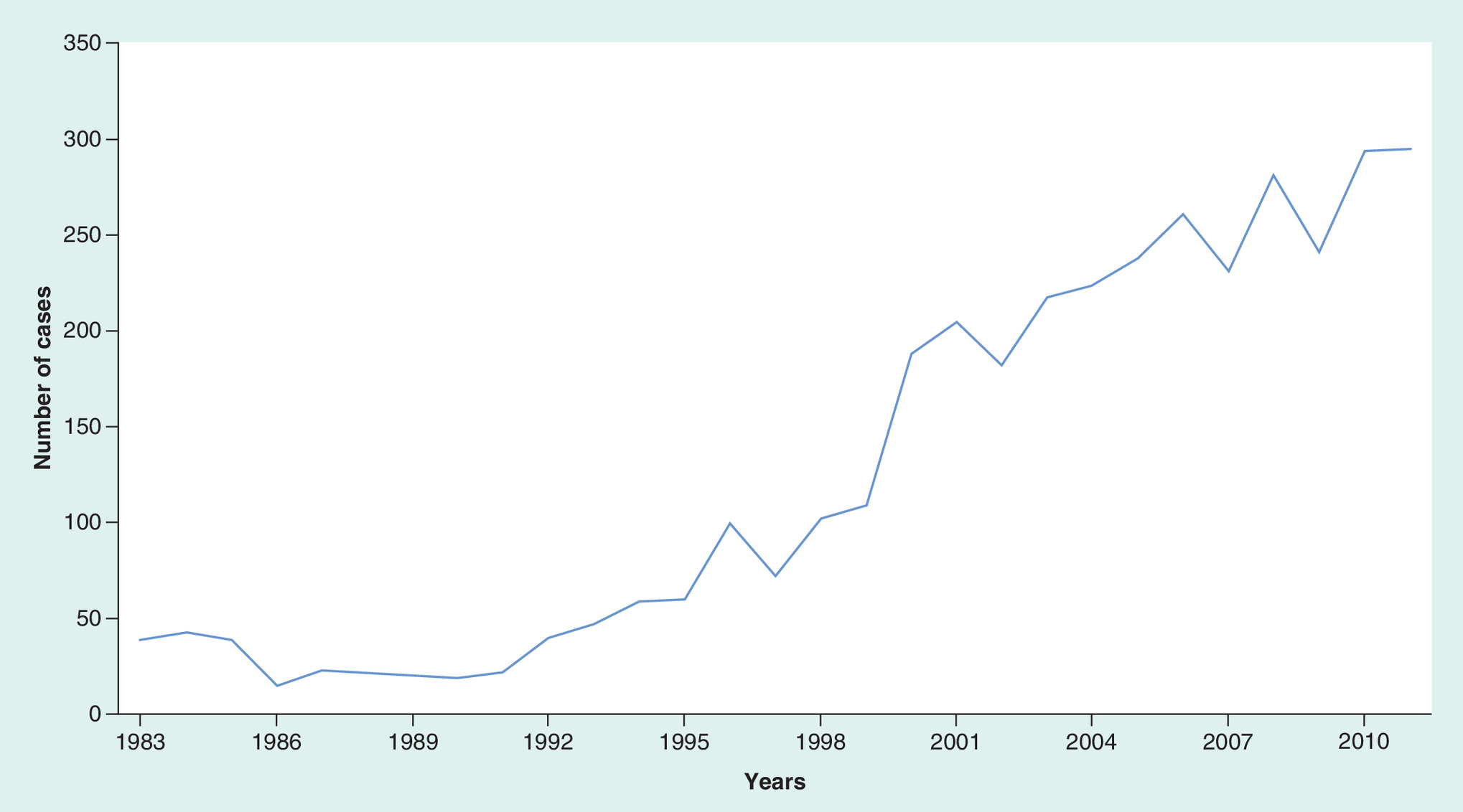
Furthermore, most patients had MCL in multiple regions, (2654, 71.9%), followed by the head, face and neck, (441, 12%) while the intrathoracic group had the least frequency (54, 1.5%). Surprisingly, there was a significantly increased incidence of NMCL cases over years with 3070 cases (83.2%) of them occurred between 1998 and 2011 as compared with 621 cases (16.8%) occurred between 1983 and 1997 (Figure 1).
There were older and more white patients in the intrathoracic group with no significant difference. Moreover, there were more patients above 50 years old in the aforementioned group, nevertheless, there were more patients ≤50 years old in the head, face and neck group. There was a significantly increased incidence of intrathoracic NMCL between 1998 and 2011. Furthermore, there were more men in the multiple regions group while there were more women in pelvic, inguinal and leg group with no significant difference. Stage I was significantly more predominant in pelvic, inguinal and leg group (67, 46.9%) than other groups while stage IV was more predominant in multiple regions group (1838, 69.3%; p < 0.001). Noteworthy, there was only one patient in stage I from multiple regions group (Supplementary Table 2). Additionally, there were more married patients in the axilla and arm group, however, there was no significant difference. There were significantly more patients who received radiation as well as had surgery in the head, face and neck group. Noteworthy, patients having multiple regions NMCL have undergone surgery less than other groups while patients with intra-abdominal MCL have received radiation less than other groups (Table 1 & Supplementary Table 3).
| Predictor | Overall (n = 3, 691) | Head, face and neck (n = 441) | Axilla and arm (n = 111) | Intrathoracic (n = 57) | Intra-abdominal (n = 285) | Pelvic, inguinal and leg (n = 143)† | Multiple regions (n = 2, 654) | p-value‡ |
|---|---|---|---|---|---|---|---|---|
| Age | 65 (12.5) | 64.1 (13.4) | 66.2 (12.1) | 70.1 (12.9) | 67.2 (11.9) | 67.5 (12) | 64.6 (12.4) | 0.68 |
| Age groups | ||||||||
| – ≤50 | 477 (12.9) | 71 (16.1) | 11 (9.9) | 5 (8.8) | 26 (9.1) | 14 (9.8) | 350 (13.2) | 0.056 |
| – >50 | 3,214 (87.1) | 370 (83.9) | 100 (90.1) | 52 (91.2) | 259 (90.9) | 129 (90.2) | 2,304 (86.8) | |
| Diagnosis year# | 2004 (8) | 2004 (9) | 2002 (10.5) | 2005 (7) | 2003 (9) | 2004 (8) | 2005 (8) | <0.001 |
| Diagnosis year groups | ||||||||
| – 1983–1997 | 621 (16.8) | 91 (20.6) | 31 (27.9) | 7 (12.3) | 70 (24.6) | 20 (14) | 402 (15.2) | <0.001 |
| – 1998–2011 | 3,070 (83.2) | 350 (79.4) | 80 (72.1) | 50 (87.7) | 215 (75.4) | 123 (86) | 2252 (84.9) | |
| Gender, male | 2567 (69.5) | 295 (66.9) | 74 (66.7) | 37 (64.9) | 184 (64.6) | 69 (48.3) | 1881 (70.9) | 0.134 |
| Race | ||||||||
| – White | 3355 (90.9) | 398 (90.2) | 101 (91) | 53 (93) | 262 (91.9) | 131 (91.6) | 2410 (90.8) | 0.66 |
| – Black | 169 (4.6) | 22 (5) | 8 (7.2) | 1 (1.8) | 9 (3.3) | 4 (2.8) | 125 (4.7) | |
| – American Indian/Alaska native | 18 (0.5) | 2 (0.5) | 0 (0) | 1 (1.8) | 2 (0.7) | 2 (1.4) | 11 (0.4) | |
| – Asian or Pacific Islander | 149 (4) | 19 (4.3) | 2 (1.8) | 2 (3.5) | 12 (4.2) | 6 (4.2) | 108 (4.1) | |
| Stage | ||||||||
| – I | 358 (9.7) | 178 (40.4) | 36 (32.4) | 15 (26.3) | 61 (21.4) | 67 (46.9) | 1 (0.04) | <0.001 |
| – II | 336 (9.1) | 84 (19) | 13 (11.7) | 11 (19.3) | 41 (14.4) | 17 (11.99) | 170 (6.4) | |
| – III | 712 (19.3) | 28 (6.3) | 12 (10.8) | 3 (5.3) | 12 (4.2) | 12 (8.4) | 645 (24.5) | |
| – IV | 2,285 (61.9) | 151 (34.2) | 50 (45) | 28 (49.1) | 171 (60) | 47 (32.9) | 1,838 (69.3) | |
| Marital status, married | 2493 (67.5) | 294 (66.7) | 79 (71.2) | 35 (61.4) | 188 (66) | 94 (65.7) | 1803 (67.9) | 0.773 |
| Survival months | 52.9 (49.7) | 66.2 (53.9) | 71.2 (67.8) | 38.1 (36.1) | 55.7 (52.3) | 57.2 (54.5) | 49.7 (47.2) | <0.001 |
| Survival rate, %§ | 29.2/42.9 | 35.4/50.8 | 32.4/54.1 | 15.8/49.1 | 25.3/39 | 31.5/51.1 | 28.6/41 | 0.005/<0.001 |
| 5-year survival rate, %¶ | 52.5 | 63 | 64.9 | 54.4 | 50.9 | 57.3 | 50.1 | <0.001 |
| 10-year survival rate, %¶ | 44.3 | 53.3 | 58.6 | 49.1 | 41.1 | 52.5 | 42.1 | <0.001 |
| Radiation, received | 385 (10.4) | 99 (22.4) | 13 (11.7) | 10 (17.5) | 14 (4.9) | 20 (14) | 229 (8.6) | <0.001 |
| Surgery, received | 1340 (36.3) | 226 (51.2) | 52 (46.8) | 15 (26.3) | 114 (40) | 57 (39.9) | 876 (24) | <0.001 |
Significant differences are in bold. Data are presented as mean (SD) for continuous variables and as frequency (percent) for categorical variables.
†
Patients with pelvic LNs were combined into inguinal region and leg LNs group because they formed of six patients only.
‡
p-value of comparison between different nodal sites.
§
The survival rate is for overall and cancer-specific survivals, respectively.
¶
Survival rate is for cancer-specific survival.
#
Data are in median and IQR.
IQR: Interaquartile range; LN: Lymph node; SD: Standard deviation.
Survival analysis
In general, the mean (standard deviation) survival was 52.9 (49.7) months with a low OS rate of 29.2% and a cancer-specific survival rate of 42.9%. In addition to that, patients having LNs involvement of the head, face and neck had significantly better survival with a mean of 66.2 (53.9) months and OS/cancer-specific survival of 35.4/50.8%, respectively, while intrathoracic-involved patients had the least survival, with a mean of 38.1 (36.1) months and OS/cancer-specific survival of 15.8/49.1% (p < 0.05; Figures 2 & 3).
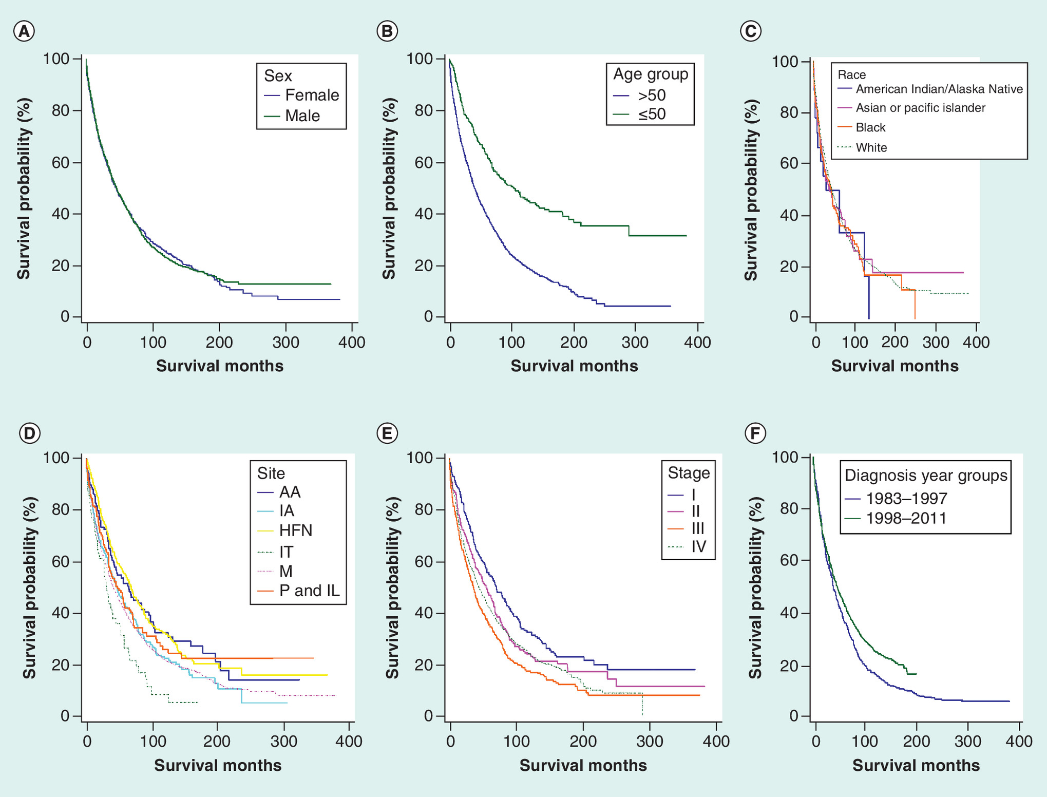
Figure 2. The Kaplan–Meier survival curves showing the overall survival for nodal mantle cell lymphoma patients.
(A) sex; (B) age; (C) race; (D) site; (E) stage; (F) diagnosis year.
KM curves revealed significantly higher OS rates in married patients, ≤50 years old, stage I, diagnosed between 1998 and 2011, received radiation and/or surgery (p < 0.05). Similar trends were found in KM curves of the cancer-specific survival (Supplementary Table 1, Figures 2 & 3 and Supplementary Table 1 & 2).

Figure 3. The Kaplan–Meier survival curves showing the cancer-specific survival for nodal mantle cell lymphoma patients.
(A) sex; (B) age; (C) race; (D) site; (E) stage; (F) diagnosis year.
Multi-variable Cox proportional hazard models
Consistently, older age, unmarried, male patients, recently diagnosed with NMCL and stage IV as compared with stages I and II were associated with a worse survival across all models (Tables 2 & 3). Moreover, American Indian/Alaska native patients had about 220% probability of more risk of overall death as compared with white patients with HR, 95% CI: 2.21, (1.21, 4.03), p = 0.01 (Table 2). In addition to that, not having surgery had a worse 5-year cancer-specific survival with HR, 95% CI: 0.86, (0.76, 0.97), p = 0.014 (Table 3).
| Predictor | Overall survival | Cancer-specific survival | ||||||
|---|---|---|---|---|---|---|---|---|
| UV Cox | MV Cox | UV Cox | MV Cox | |||||
| HR, 95% CI | p-value | HR, 95% CI | p-value | HR, 95% CI | p-value | HR, 95% CI | p-value | |
| Age, years | 1.04 (1.04, 1.05) | <0.001 | 1.04 (1.04, 1.05) | <0.001 | 1.04 (1.03, 1.04) | <0.001 | 1.04 (1.03, 1.04) | <0.001 |
| Year of diagnosis | 0.98 (0.98, 0.99) | <0.001 | 0.98 (0.97, 0.98) | <0.001 | 0.98 (0.98, 0.99) | <0.001 | 0.98 (0.97, 0.99) | <0.001 |
| Gender | ||||||||
| – Male | Reference | |||||||
| – Female | 0.998 (0.92, 1.08) | 0.955 | 0.82 (0.74, 0.91) | <0.001 | 0.98 (0.89, 1.07) | 0.649 | 0.85 (0.76, 0.96) | 0.006 |
| Race | ||||||||
| – White | Reference | |||||||
| – Black | 1.05 (0.88, 1.26) | 0.59 | 1.14 (0.92, 1.41) | 0.231 | 1 (0.81, 1.23) | 0.985 | 1.06 (0.83, 1.36) | 0.622 |
| – American Indian/Alaska native | 1.13 (0.66, 1.95) | 0.659 | 2.21 (1.21, 4.03) | 0.01 | 0.96 (0.496, 1.84) | 0.89 | 1.98 (0.98, 3.98) | 0.057 |
| – Asian or Pacific Islander | 1.03 (0.84, 1.25) | 0.795 | 1.23 (0.97, 1.56) | 0.09 | 0.91 (0.72, 1.15) | 0.445 | 1.09 (0.82, 1.44) | 0.55 |
| Primary site | ||||||||
| – Axilla and arm | Reference | |||||||
| – Intra-abdominal | 1.31 (1.004, 1.7) | 0.047 | 1.29 (0.94, 1.75) | 0.112 | 1.54 (1.13, 2.1) | 0.007 | 1.60 (1.102, 2.33) | 0.013 |
| – Head, face and neck | 0.97 (0.75, 1.25) | 0.821 | 1.23 (0.91, 1.66) | 0.172 | 1.07 (0.79, 1.45) | 0.665 | 1.46 (1.01, 2.1) | 0.043 |
| – Pelvic, inguinal and leg† | 1.18 (0.87, 1.59) | 0.288 | 1.36 (0.95, 1.95) | 0.096 | 1.21 (0.84, 1.74) | 0.296 | 1.61 (1.05, 2.49) | 0.031 |
| – Multiple regions | 1.35 (1.07, 1.7) | 0.01 | 1.402 (1.06, 1.85) | 0.018 | 1.6 (1.21, 2.11) | 0.001 | 1.61 (1.14, 2.27) | 0.006 |
| – Intrathoracic | 1.93 (1.35, 2.78) | <0.001 | 1.796 (1.17, 2.76) | 0.007 | 1.64 (1.04, 2.59) | 0.034 | 1.37 (0.77, 2.42) | 0.281 |
| Stage | ||||||||
| – I | 0.73 (0.63, 0.83) | <0.001 | 0.61 (0.504, 0.75) | <0.001 | 0.55 (0.46, 0.65) | <0.001 | 0.48 (0.38, 0.61) | <0.001 |
| – II | 0.92 (0.802, 1.05) | 0.228 | 0.79 (0.67, 0.93) | 0.006 | 0.83 (0.71, 0.97) | 0.021 | 0.77 (0.64, 0.92) | <0.001 |
| – III | 1.2 (1.09, 1.32) | <0.001 | 0.99 (0.88, 1.12) | 0.919 | 1.16 (1.04, 1.29) | 0.006 | 0.96 (0.84, 1.1) | 0.549 |
| – IV | Reference | |||||||
| Marital status | ||||||||
| – Married | Reference | |||||||
| – Not married | 1.33 (1.22, 1.44) | <0.001 | 1.3 (1.18, 1.44) | <0.001 | 1.24 (1.13, 1.36) | <0.001 | 1.23 (1.1, 1.37) | <0.001 |
| Radiation | ||||||||
| – None/unknown | Reference | |||||||
| – Received radiation | 0.78 (0.69, 0.89) | <0.001 | 1.05 (0.903, 1.23) | 0.502 | 0.77 (0.66, 0.89) | <0.001 | 1.08 (0.91, 1.29) | 0.355 |
| Surgery | ||||||||
| – No surgery | Reference | |||||||
| – Received surgery | 0.795 (0.73, 0.86) | <.001 | 0.93 (0.84, 1.03) | 0.158 | 0.77 (0.699, 0.84) | <0.001 | 0.91 (0.82, 1.02) | 0.106 |
Significant predictors are in bold.
†
Patients with pelvic LNs were combined into inguinal region and leg LNs because they were formed of six patients only.
HR: Hazard ratio; LN: Lymph node; MV: Multivariable; UV: Univariable.
| Predictor | 5-year cancer-specific survival | 10-year cancer-specific survival | ||
|---|---|---|---|---|
| HR, 95% CI | p-value | HR, 95% CI | p-value | |
| Age, years | 1.04 (1.03, 1.04) | <0.001 | 1.04 (1.03, 1.04) | <0.001 |
| Year of diagnosis | 0.98 (0.98, 0.99) | <0.001 | 0.98 (0.97, 0.99) | <0.001 |
| Gender | ||||
| – Male | Reference | |||
| – Female | 0.87 (0.77, 0.99) | 0.029 | 0.84 (0.75, 0.95) | 0.004 |
| Race | ||||
| – White | Reference | |||
| – Black | 1.07 (0.82, 1.4) | 0.605 | 1.04 (0.81, 1.34) | 0.739 |
| – American Indian/Alaska native | 1.64 (0.73, 3.68) | 0.23 | 1.7 (0.81, 3.6) | 0.163 |
| – Asian or Pacific Islander | 1.08 (0.8, 1.46) | 0.63 | 1.11 (0.84, 1.47) | 0.449 |
| Primary site | ||||
| – Axilla and arm | Reference | |||
| – Intra-abdominal | 1.63 (1.07, 2.5) | 0.024 | 1.66 (1.12, 2.46) | 0.011 |
| – Head, face and neck | 1.32 (0.87, 2.01) | 0.194 | 1.47 (0.998, 2.15) | 0.051 |
| – Pelvic, inguinal and leg† | 1.67 (1.03, 2.7) | 0.039 | 1.64 (1.04, 2.58) | 0.032 |
| – Multiple regions | 1.61 (1.09, 2.39) | 0.017 | 1.69 (1.18, 2.42) | 0.005 |
| – Intrathoracic | 1.36 (0.74, 2.51) | 0.322 | 1.43 (0.8, 2.56) | 0.23 |
| Stage | ||||
| – I | 0.51 (0.4, 0.67) | <0.001 | 0.51 (0.397, 0.65) | <0.001 |
| – II | 0.76 (0.62, 0.94) | 0.011 | 0.8 (0.67, 0.97) | 0.021 |
| – III | 0.92 (0.795, 1.06) | 0.256 | 0.97 (0.85, 1.11) | 0.637 |
| – IV | Reference | |||
| Marital status | ||||
| – Married | Reference | |||
| – Not married | 1.3 (1.15, 1.47) | <0.001 | 1.25 (1.12, 1.4) | <0.001 |
| Radiation | ||||
| – No/unknown | Reference | |||
| – Received radiation | 1.07 (0.88, 1.29) | 0.498 | 1.08 (0.904, 1.28) | 0.409 |
| Surgery | ||||
| – No surgery | Reference | |||
| – Received surgery | 0.86 (0.76, 0.97) | 0.014 | 0.91 (0.82, 1.02) | 0.114 |
Significant predictors are in bold.
†
Patients with pelvic LNs were combined into inguinal region and leg LNs because they were formed of six patients only.
HR: Hazard ratio; LN: Lymph node.
For the primary site of NMCL, intrathoracic and multiple regions groups had a worse OS as compared with the axilla and arm group with 40 and 79% probability of more risk of overall death, respectively, while the head, face and neck (46%), intra-abdominal (60%), pelvic, inguinal and leg (61%) and multiple regions (61%) had a worse cancer-specific survival (Table 2). Consistent with the cancer-specific survival, the 5- and 10-year cancer-specific survival had similar trends (Table 3).
The Cox prediction models validation
Our models had an accuracy ranged between 63.3 and 72.6% with an area under the curve of 0.67–0.75, a sensitivity of 58.6–66.3% and a specificity of 61.8–76.5% (Supplementary Table 4).
Discussion
Our current study has disclosed several factors, including age, sex, primary site, marital status, race, stage, diagnosis year and surgery influencing the survival in NMCL patients.
It was revealed that older patients had worse survival. Indeed, they are most likely to have associated comorbidities that limit the therapeutic choices. Moreover, allogeneic hematopoietic stem cells transplant is offered almost exclusively to younger patients [40,41]. Furthermore, the advanced age is associated with worse outcomes in all subsets of NHL [42,43]. Akin to that, Cohen et al. and Zhou et al. magnify a good survival in patients who are <60 and <50 years old, respectively [11,44].
Men had lower survival than women, this substantiates previous results of Chandran et al. which highlight a higher MCL mortality in men and supports our finding that sex plays an important role in controlling survival in MCL [13]. It may be due to the hormonal differences that result in promising tumor characteristics [45] and a better response to immune-chemotherapy (rituximab) since this drug has been comprehensively used in MCL [46].
On the other hand, stage IV had the worst survival across all models, similarly advanced stages (III/IV) were found to be independently associated with increased mortality [18,47, 54-56]. Murthy et al. included stage I and II patients, have found better survival in the former [49].
In addition to that, married patients had a better survival as well, this supports previous findings in other cancers [48,50–56]. Also, that discloses the optimal and significant influence that marriage, especially social support, will have on cancer detection, treatment and even survival. Aizer et al. found that unmarried patients, including those who are widowed, are at a significantly greater risk of the exhibition with metastatic cancer, under treatment and even mortality from their cancer than married patients. After adjusting for other confounders, the marriage remained associated with a decline in the mortality ranging from 12 to 33% [52,53].
We could not find that receiving the radiation reduces mortality. This may be due to the small number of patients received radiation in our study (385, 10.4%) with similar insignificant results in Ambinder et al. study (307, 8.7%) [18].
Noteworthy, we have revealed that patients who underwent surgery had a better survival across all models, however, it was only significant in 5-year cancer-specific survival and other univariable but not other multivariable analyses. Interestingly, patients who underwent surgery had better survival rates than who did not undergo, 91.5% compared with 68.8%, respectively [57]. Undeniably, performing surgery in some sites such as GI lymphomas has a perfect impact on patients’ survival and in confirming a correct pathological staging and a precise histological diagnosis [57–70]. MCL treatment varies from chemotherapy to autologous stem cell transplantation with contrary results varying from good to a bad survival rate [7,71–73]. The resistance to chemotherapy marks an aggressive incurable disease requiring special attention from clinicians [37,74–77].
We have revealed an increased incidence of NMCL. Moreover, several studies have reported increasing the trends in the US [10,12–14]. Recent studies suggested an improved survival in the non-nodal type of MCL [33–36]. Furthermore, NMCL is more common than extra-nodal MCL. Notwithstanding, the worsening in the survival of NMCL in this study is probably because most of the patients were male, in stage IV with a mean age of 65 years and the majority was above 50 years old, in addition to that there was a predominance of LNs involvement of multiple regions. A previous study has revealed a worsening in the survival of NMCL as compared with the extra-nodal involvement with 30% probability of more risk of death [51].
In addition, the disease primary site can serve as an essential prognostic factor for MCL patients’ survival. Although MCL characteristics, survival and treatment were discussed in previous studies, MCL is still an unresolved problem among clinicians. An invasion of the GI tract occurs in the advanced stage of the disease, however, primary GI MCL rarely occurs with an incidence of 2% of primary GI lymphoma [78–80]. The primary site of the disease was demonstrated before as an essential and accurate prognostic factor in other cancers such as diffuse large B-cell lymphoma [81,82].
We have found several primary sites associated with increased mortality. NMCL of multiple regions involved LNs was on the top of them followed by intrathoracic, the head, face and neck, intra-abdominal and pelvic, inguinal and leg as well. Of note, patients with multiple regions involved LNs have undergone surgery less than other groups with more males than females and a significant stage IV predominance with only one patient in stage I. Similarly, there were more married patients in the axilla and arm group that may explain their better survival. Also, there was a significantly increased incidence of intrathoracic NMCL between 1998 and 2011. Indeed, there were more patients above 50 years old in that aforementioned group.
A biological mechanism should be disclosed for these primary sites aggressiveness. Undoubtedly, the rising belief of a clonal similarity among complex lymphomas different components, including mucosa-associated lymphoid tissue lymphoma and follicular lymphoma (FL) or Hodgkin’s lymphoma (HL) and FL or MCL, paves the way to simplify the complex appearance of these tumors, which may share another subtler but a common tumor behavior [83].
It has been demonstrated that a low-grade FL could be transformed into a highly aggressive large B-cell lymphoma and that histologic transformation could be clinically associated with the abrupt explosive growth of a single LN site, leading to a median survival time of 6–20 months only [25,26]. Subsequently, the resulting composite lymphoma will have the same lineage, yet other composite lymphomas were described to have different lineages (B and T cells) or compromising HL, as well as NHL combinations [27].
Regarding the primary LNs site influence on the survival in NMCL patients, several clinicopathologic characteristics of patients with these aforementioned primary sites should be investigated to see if similarities are present that may indicate either resemblance or transformation to a more aggressive or indolent tumor type.
Thus, we strongly encourage future studies to compare NMCL patients’ survival affected by the aggressive primary sites with other unfavorable variables in MIPI. Subsequently, we strongly encourage adding the primary site, especially, the multiple NMCL in the MIPI.
It has not been investigated before the association between the primary site and NMCL. Notwithstanding, we had some limitations including its retrospective nature and quite low accuracy, sensitivity and specificity of the prediction models. However, with that large sample size and several prediction models including OS, cancer-specific survival and 5- and 10-year cancer-specific survival, these limitations may not have effects on the current analysis with nearly the same significant predictors in all prediction models. Moreover, we could not validate our new prognostic factors against the MIPI since SEER does not have this index, thus future studies need to validate our findings against the MIPI.
Conclusion
NMCL has a worse survival with an increased incidence over the years. Several predictors have been identified. Older age, unmarried, male patients, recently diagnosed with MCL and stage IV were associated with worse survival. Multiple regions, intrathoracic, the head, face and neck, intra-abdominal, as well as pelvic, inguinal and leg LNs were associated with a worse survival while axilla and arm LNs were associated with better survival, suggesting that NMCL primary site can serve as a prognostic factor. Further studies need to investigate the biological basis of these aggressive primary sites and to validate its accuracy then it is encouraged to be added to MIPI.
•
Nodal mantle cell lymphoma (NMCL) has a worse survival than extra-nodal MCL.
•
NMCL had an increased incidence over the years with a mean survival of 52.9 months and overall survival/cancer-specific survival rate of 29.2/42.9%, respectively.
•
Older age, unmarried, male patients, recently diagnosed with mantle cell lymphoma and stage IV were associated with a worse survival.
•
Multiple-involved regions, intrathoracic, the head, face and neck, intra-abdominal, as well as pelvic, inguinal region and leg lymph nodes (LNs) were associated with a worse survival while axilla and arm LNs were associated with a better survival.
•
NMCL primary site can serve as a prognostic factor.
•
Further studies need to investigate the biological basis of these aggressive primary sites and to validate its accuracy then it is encouraged to be added to MCL International Prognostic Index.
Financial & competing interests disclosure
The authors have no relevant affiliations or financial involvement with any organization or entity with a financial interest in or financial conflict with the subject matter or materials discussed in the manuscript. This includes employment, consultancies, honoraria, stock ownership or options, expert testimony, grants or patents received or pending, or royalties.
No writing assistance was utilized in the production of this manuscript.
Supplementary Material
File (suppl_file.docx)
- Download
- 191.89 KB
References
Papers of special note have been highlighted as: • of interest; •• of considerable interest
1.
Harris NL, Jaffe ES, Stein H et al. A revised European-American classification of lymphoid neoplasms: a proposal from the International Lymphoma Study Group. Blood 84(5), 1361–1392 (1994).
2.
Harris NL, Jaffe ES, Diebold J et al. World Health Organization classification of neoplastic diseases of the hematopoietic and lymphoid tissues: report of the Clinical Advisory Committee meeting – Airlie House, Virginia, November 1997. J. Clin. Oncol. 17(12), 3835–3849 (1999).
3.
Samaha H, Dumontet C, Ketterer N et al. Mantle cell lymphoma: a retrospective study of 121 cases. Leukemia 12(8), 1281–1287 (1998).
4.
Mannina D, Loteta B. Ibrutinib treatment of mantle cell lymphoma relapsing at central nervous system: a case report and literature review. Case Rep. Hematol. 2017, 9583257 (2017).
5.
Cheah CY, George A, Gine E et al. Central nervous system involvement in mantle cell lymphoma: clinical features, prognostic factors and outcomes from the European Mantle Cell Lymphoma Network. Ann. Oncol. 24(8), 2119–2123 (2013).
6.
Ferrer A, Bosch F, Villamor N et al. Central nervous system involvement in mantle cell lymphoma. Ann. Oncol. 19(1), 135–141 (2008).
7.
Dreyling M, Hiddemann W. Current treatment standards and emerging strategies in mantle cell lymphoma. In: ASH Education Program Book 2009(1), 542–551 (2009).
8.
Fernandez V, Salamero O, Espinet B et al. Genomic and gene expression profiling defines indolent forms of mantle cell lymphoma. Cancer Res. 70(4), 1408–1418 (2010).
9.
Bosch F, López-Guillermo A, Campo E et al. Mantle cell lymphoma. Cancer 82(3), 567–575 (1998).
10.
Morton LM, Wang SS, Devesa SS, Hartge P, Weisenburger DD, Linet MS. Lymphoma incidence patterns by WHO subtype in the United States, 1992-2001. Blood 107(1), 265–276 (2006).
11.
Zhou Y, Wang H, Fang W et al. Incidence trends of mantle cell lymphoma in the United States between 1992 and 2004. Cancer 113(4), 791–798 (2008).
12.
Shiels MS, Engels EA, Linet MS et al. The epidemic of non-Hodgkin lymphoma in the United States: disentangling the effect of HIV, 1992-2009. Cancer Epidemiol. Prev. Biomark. 22(6), 1069–1078 (2013).
13.
Chandran R, Gardiner SK, Simon M, Spurgeon SE. Survival trends in mantle cell lymphoma in the United States over 16 years 1992-2007. Leuk. Lymphoma 53(8), 1488–1493 (2012).
14.
Zhou Y, Wang H, Fang W et al. Incidence trends of mantle cell lymphoma in the United States between 1992 and 2004. Cancer 113(4), 791–798 (2008).
15.
Kluin-Nelemans H, Hoster E, Hermine O et al. Treatment of older patients with mantle-cell lymphoma. N. Engl. J. Med. 367(6), 520–531 (2012).
16.
Campo E, Raffeld M, Jaffe ES. Mantle-cell lymphoma. Semin. Hematol. 36(2), 115–127 (1999).
17.
Swerdlow SH, Williams ME. From centrocytic to mantle cell lymphoma: a clinicopathologic and molecular review of 3 decades. Human Pathol. 33(1), 7–20 (2002).
18.
Ambinder AJ, Shenoy PJ, Nastoupil LJ, Flowers CR. Using primary site as a predictor of survival in mantle cell lymphoma. Cancer 119(8), 1570–1577 (2013).
•• A study investigating the primary site role in extranodal mantle cell lymphoma.
19.
Rosenwald A, Wright G, Wiestner A et al. The proliferation gene expression signature is a quantitative integrator of oncogenic events that predicts survival in mantle cell lymphoma. Cancer Cell 3(2), 185–197 (2003).
20.
Espinet B, Solé F, Pedro C et al. Clonal proliferation of cyclin D1-positive mantle lymphocytes in an asymptomatic patient: an early-stage event in the development or an indolent form of a mantle cell lymphoma? Human Pathol. 36(11), 1232–1237 (2005).
21.
Martin P, Chadburn A, Christos P et al. Outcome of deferred initial therapy in mantle-cell lymphoma. J. Clin. Oncol. 27(8), 1209–1213 (2009).
22.
Nodit L, Bahler DW, Jacobs SA, Locker J, Swerdlow SH. Indolent mantle cell lymphoma with nodal involvement and mutated immunoglobulin heavy chain genes. Human Pathol. 34(10), 1030–1034 (2003).
23.
Orchard J, Garand R, Davis Z et al. A subset of t(11;14) lymphoma with mantle cell features displays mutated IgVH genes and includes patients with good prognosis, nonnodal disease. Blood 101(12), 4975–4981 (2003).
24.
Sander B. Mantle cell lymphoma: recent insights into pathogenesis, clinical variability, and new diagnostic markers. Semin. Diagn. Pathol. 28(3), 245–255 (2011).
25.
Hoster E, Dreyling M, Klapper W et al. A new prognostic index (MIPI) for patients with advanced-stage mantle cell lymphoma. Blood 111(2), 558–565 (2008).
26.
Van De Schans SA, Janssen-Heijnen ML, Nijziel MR, Steyerberg EW, Van Spronsen DJ. Validation, revision and extension of the Mantle Cell Lymphoma International Prognostic Index in a population-based setting. Haematologica 95(9), 1503–1509 (2010).
27.
Todorovic M, Balint B, Andjelic B et al. Outcome prediction of advanced mantle cell lymphoma by international prognostic index versus different mantle cell lymphoma indexes: one institution study. Med. Oncol. 29(3), 2212–2219 (2012).
28.
Geisler CH, Kolstad A, Laurell A et al. The Mantle Cell Lymphoma International Prognostic Index (MIPI) is superior to the International Prognostic Index (IPI) in predicting survival following intensive first-line immunochemotherapy and autologous stem cell transplantation (ASCT). Blood 115(8), 1530–1533 (2010).
29.
Hoster E. Prognostic relevance of clinical risk factors in mantle cell lymphoma. Semin. Hematol. 48(3), 185–188 (2011).
30.
Determann O, Hoster E, Ott G et al. Ki-67 predicts outcome in advanced-stage mantle cell lymphoma patients treated with anti-CD20 immunochemotherapy: results from randomized trials of the European MCL Network and the German Low Grade Lymphoma Study Group. Blood 111(4), 2385–2387 (2008).
31.
Greiner TC, Moynihan MJ, Chan WC et al. p53 mutations in mantle cell lymphoma are associated with variant cytology and predict a poor prognosis. Blood 87(10), 4302–4310 (1996).
32.
Hernandez L, Fest T, Cazorla M et al. p53 gene mutations and protein overexpression are associated with aggressive variants of mantle cell lymphomas. Blood 87(8), 3351–3359 (1996).
33.
Orchard J, Garand R, Davis Z et al. A subset of t (11; 14) lymphoma with mantle cell features displays mutated IgV H genes and includes patients with good prognosis, nonnodal disease. Blood 101(12), 4975–4981 (2003).
34.
Fernàndez V, Salamero O, Espinet B et al. Genomic and gene expression profiling defines indolent forms of mantle cell lymphoma. Cancer Res. 70(4), 1408–1418 (2010).
35.
Ondrejka SL, Lai R, Smith SD, Hsi ED. Indolent mantle cell leukemia: a clinicopathological variant characterized by isolated lymphocytosis, interstitial bone marrow involvement, kappa light chain restriction, and good prognosis. Haematologica 96(8), 1121–1127 (2011).
36.
Furtado M, Rule S. Indolent mantle cell lymphoma. Haematologica 96(8), 1086–1088 (2011).
37.
Jurczak W, Ramanathan S, Giri P et al. Comparison of two doses of intravenous temsirolimus in patients with relapsed/refractory mantle cell lymphoma. Leuk. Lymphoma 59(3), 670–678 (2018).
38.
Turner JJ, Morton LM, Linet MS et al. InterLymph hierarchical classification of lymphoid neoplasms for epidemiologic research based on the WHO classification (2008): update and future directions. Blood 116(20), e90–e98 (2010).
39.
Morton LM, Turner JJ, Cerhan JR et al. Proposed classification of lymphoid neoplasms for epidemiologic research from the Pathology Working Group of the International Lymphoma Epidemiology Consortium (InterLymph). Blood 110(2), 695–708 (2007).
40.
Andersen N, Jensen M, De Nully Brown P, Geisler C. A Danish population-based analysis of 105 mantle cell lymphoma patients: incidences, clinical features, response, survival and prognostic factors. Eur. J. Cancer 38(3), 401–408 (2002).
• A Danish population-based study of mantle cell lymphoma investigating incidences, clinical features, response, survival and prognostic factors.
41.
Velders G, Kluin-Nelemans J, De Boer C et al. Mantle-cell lymphoma: a population-based clinical study. J. Clin. Oncol. 14(4), 1269–1274 (1996).
42.
Vose JM, Armitage J, Weisenburger D et al. The importance of age in survival of patients treated with chemotherapy for aggressive non-Hodgkin’s lymphoma. J. Clin. Oncol. 6(12), 1838–1844 (1988).
43.
Armitage JO, Potter JF. Aggressive chemotherapy for diffuse histiocytic lymphoma in the elderly: increased complications with advancing age. J. Am. Geriatrics Soc. 32(4), 269–273 (1984).
44.
Cohen JB, Han X, Jemal A, Ward EM, Flowers CR. Deferred therapy is associated with improved overall survival in patients with newly diagnosed mantle cell lymphoma. Cancer 122(15), 2356–2363 (2016).
45.
Pal SK, Hurria A. Impact of age, sex, and comorbidity on cancer therapy and disease progression. J. Clin. Oncol. 28(26), 4086–4093 (2010).
46.
Riihijärvi S, Taskinen M, Jerkeman M, Leppä S. Male gender is an adverse prognostic factor in B-cell lymphoma patients treated with immunochemotherapy. Eur. J. Haematol. 86(2), 124–128 (2011).
47.
Kamel MG, Istanbuly S, Abd-Elhay Fa-E et al. Examined and positive lymph node counts are associated with mortality in prostate cancer: a population-based analysis. Urol. Int. (2020) (Epub ahead of print).
48.
Kamel MG, El-Qushayri AE, Thach TQ, Huy NT. Cardiovascular mortality trends in non-Hodgkin's lymphoma: a population-based cohort study. Expert Rev. Anticancer Ther. 18(1), 91–100 (2018).
49.
Elhusseiny KM, Abd-Elhay FA, Kamel MG et al. Examined and positive lymph nodes counts and lymph nodes ratio are associated with survival in major salivary gland cancer. Head Neck 41(8), 2625–2635 (2019).
50.
Abd-Elhay FA, Elhusseiny KM, Kamel MG et al. Negative lymph node count and lymph node ratio are associated with survival in male breast cancer. Clin. Breast Cancer 18(6), e1293–e1310 (2018).
51.
Guru Murthy GS, Venkitachalam R, Mehta P. Effect of radiotherapy on the survival of patients with stage I and stage II mantle cell lymphoma: analysis of the Surveillance, Epidemiology and End Results database. Clin. Lymphoma Myeloma Leuk. 14(Suppl.), S90–S95 (2014).
52.
Cronin-Fenton DP, Sharp L, Deady S, Comber H. Treatment and survival for non-Hodgkin’s lymphoma: influence of histological subtype, age, and other factors in a population-based study (1999-2001). Eur. J. Cancer 42(16), 2786–2793 (2006).
53.
Ross R, Dworsky R, Paganini-Hill A, Levine A, Mack T. Non-Hodgkin’s lymphomas in never married men in Los Angeles. Brit. J. Cancer 52(5), 785 (1985).
54.
Aizer AA, Chen M-H, Mccarthy EP et al. Marital status and survival in patients with cancer. J. Clin. Oncol. 31(31), 3869–3876 (2013).
55.
Abouyabis AN, Shenoy PJ, Lechowicz MJ, Flowers CR. Incidence and outcomes of the peripheral T-cell lymphoma subtypes in the United States. Leuk. Lymphoma 49(11), 2099–2107 (2008).
56.
Koch P, Del Valle F, Berdel WE et al. Primary gastrointestinal non-Hodgkin’s lymphoma: II. Combined surgical and conservative or conservative management only in localized gastric lymphoma-results of the prospective German Multicenter Study GIT NHL 01/92. J. Clin. Oncol. 19(18), 3874–3883 (2001).
57.
Gospodarowicz MK, Bush R, Brown J, Chua T. Curability of gastrointestinal lymphoma with combined surgery and radiation. Int. J. Radiat. Oncol. Biol. Phys. 9(1), 3–9 (1983).
58.
Fleming ID, Mitchell S, Dilawari RA. The role of surgery in the management of gastric lymphoma. Cancer 49(6), 1135–1141 (1982).
59.
Paulson S, Sheehan R, Stone M, Frenkel E. Large cell lymphomas of the stomach: improved prognosis with complete resection of all intrinsic gastrointestinal disease. J. Clin. Oncol. 1(4), 263–269 (1983).
60.
Herrera A, Solal-Celigny P, Bernard J. High-risk primary lymphomas of the digestive tract: therapeutic results and study of prognostic factors. In: Non-Hodgkin's Lymphomas: New Techniques and Treatments. Sotto JJ, Vrousos C, Sotto M-F, Vincent F (Eds). Karger Publishers 206–209 (1985).
61.
Dragosics B, Bauer P, Radaszkiewicz T. Primary gastrointestinal non-Hodgkin’s lymphomas. A retrospective clinicopathologic study of 150 cases. Cancer 55(5), 1060–1073 (1985).
62.
Rosenfelt F, Rosenberg SA. Diffuse histiocytic lymphoma presenting with gastrointestinal tract lesions the stanford experience. Cancer 45(8), 2188–2193 (1980).
63.
Ampil F. Primary gastrointestinal lymphoma. Oncology 44(4), 214–218 (1987).
64.
Economopoulos T, Alexopoulos C, Stathakis N et al. Primary gastric lymphoma – the experience of a general hospital. Brit. J. Cancer 52(3), 391 (1985).
65.
Sheridan WP, Medley G, Brodie GN. Non-Hodgkin’s lymphoma of the stomach: a prospective pilot study of surgery plus chemotherapy in early and advanced disease. J. Clin. Oncol. 3(4), 495–500 (1985).
66.
Liang R, Todd D, Chan T, Ng R, Ho F. Gastrointestinal lymphoma in Chinese: a retrospective analysis. Hematol. Oncol. 5(2), 115–126 (1987).
67.
Steward WP, Harris M, Wagstaff J et al. A prospective study of the treatment of high-grade histology non-Hodgkin’s lymphoma involving the gastrointestinal tract. Eur. J. Cancer Clin. Oncol. 21(10), 1195–1200 (1985).
68.
List AF, Greer J, Cousar J et al. Non-Hodgkin’s lymphoma of the gastrointestinal tract: an analysis of clinical and pathologic features affecting outcome. J. Clin. Oncol. 6(7), 1125–1133 (1988).
69.
Shepherd FA, Evans W, Kutas G et al. Chemotherapy following surgery for stages IE and IIE non-Hodgkin’s lymphoma of the gastrointestinal tract. J. Clin. Oncol. 6(2), 253–260 (1988).
70.
Janus C, Edwards B, Sariban E, Magrath I. Surgical resection and limited chemotherapy for abdominal undifferentiated lymphomas. Cancer Treat. Rep. 68(4), 599–605 (1984).
71.
Guidot DM, Switchenko JM, Nastoupil LJ et al. Surveillance imaging in mantle cell lymphoma in first remission lacks clinical utility. Leuk. Lymphoma 1–8 (2017) (Epub ahead of print).
72.
Geisler CH, Kolstad A, Laurell A et al. Long-term progression-free survival of mantle cell lymphoma after intensive front-line immunochemotherapy with in vivo-purged stem cell rescue: a nonrandomized Phase II multicenter study by the Nordic Lymphoma Group. Blood 112(7), 2687–2693 (2008).
73.
Kahl BS, Longo WL, Eickhoff JC et al. Maintenance rituximab following induction chemoimmunotherapy may prolong progression-free survival in mantle cell lymphoma: a pilot study from the Wisconsin Oncology Network. Ann. Oncol. 17(9), 1418–1423 (2006).
74.
Cortelazzo S, Ponzoni M, Ferreri AJ, Dreyling M. Mantle cell lymphoma. Crit. Rev. Oncol. Hematol. 82(1), 78–101 (2012).
75.
Armitage JO. A clinical evaluation of the International Lymphoma Study Group classification of non-Hodgkin’s lymphoma. Blood 89(11), 3909–3918 (1997).
76.
Tiemann M, Schrader C, Klapper W et al. Histopathology, cell proliferation indices and clinical outcome in 304 patients with mantle cell lymphoma (MCL): a clinicopathological study from the European MCL Network. Brit. J. Haematol. 131(1), 29–38 (2005).
77.
Argatoff LH, Connors JM, Klasa RJ, Horsman DE, Gascoyne RD. Mantle cell lymphoma: a clinicopathologic study of 80 cases. Blood 89(6), 2067–2078 (1997).
78.
Dasappa L, Suresh Babu MC, Sirsath NT et al. Primary gastrointestinal mantle cell lymphoma: a retrospective study. J. Gastrointest. Cancer 45(4), 481–486 (2014).
79.
Salar A, Juanpere N, Bellosillo B et al. Gastrointestinal involvement in mantle cell lymphoma: a prospective clinic, endoscopic, and pathologic study. Am. J. Surg. Pathol. 30(10), 1274–1280 (2006).
80.
Morton JE, Leyland MJ, Vaughan Hudson G et al. Primary gastrointestinal non-Hodgkin’s lymphoma: a review of 175 British National Lymphoma Investigation cases. Br. J. Cancer 67(4), 776–782 (1993).
81.
Kamel MG, Abd-Elhay FA, Morsy S et al. The role of primary lymph node sites in survival and mortality prediction in DLBCL: a SEER population-based retrospective study [abstract]. Cancer Res. 78(Suppl. 13), 2215 (2018).
•• The role of primary lymph node sites in survival and mortality prediction in diffuse large B-cell lymphoma.
82.
Castillo JJ, Winer ES, Olszewski AJ. Sites of extranodal involvement are prognostic in patients with diffuse large B-cell lymphoma in the rituximab era: an analysis of the Surveillance, Epidemiology and End Results database. Am. J. Hematol. 89(3), 310–314 (2014).
83.
Ebied A, Thanh Huan V, Makram OM et al. The role of primary lymph node sites in survival and mortality prediction in Hodgkin lymphoma: a SEER population-based retrospective study. Cancer Med. 7(4), 953–965 (2018).
• The primary site role in Hodgkin’s lymphoma patients.
84.
Swerdlow SH, Campo E, Pileri SA et al. The 2016 revision of the World Health Organization classification of lymphoid neoplasms. Blood 127(20), 2375–2390 (2016).
Information & Authors
Information
Published In
Pages: 861 - 876
PubMed: 32885984
Copyright
© 2020 Future Medicine Ltd.
History
Received: 14 May 2020
Accepted: 9 July 2020
Published online: 4 September 2020
Keywords:
Topics
Authors
Metrics & Citations
Metrics
Article Usage
Article usage data only available from February 2023. Historical article usage data, showing the number of article downloads, is available upon request.
Citations
How to Cite
Using the primary site as a prognostic tool for nodal mantle cell lymphoma: a SEER-based study. (2020) Journal of Comparative Effectiveness Research. DOI: 10.2217/cer-2020-0083
Export citation
Select the citation format you wish to export for this article or chapter.
Citing Literature
- Ting Zhang, Zhao-tong Wang, Zhuo Li, Shuo-xin Yin, Xun Wang, Hai-zhu Chen, The influence of marital status at diagnosis on survival of adult patients with mantle cell lymphoma, Journal of Cancer Research and Clinical Oncology, 10.1007/s00432-024-05647-z, 150, 3, (2024).
- Marcus Vinícius Ribeiro Carvalho, Carla Isabelly Rodrigues-Fernandes, Cinthia Veronica Bardález López de Cáceres, Ricardo Alves Mesquita, Manoela Domingues Martins, Elena María José Román Tager, Roman Carlos, Fábio Ramôa Pires, Liam Robinson, Willie F.P. van Heerden, Vitor Bonetti Valente, José Cândido Caldeira Xavier-Júnior, Daniel Galera Bernabé, Hélder Antônio Rebelo Pontes, Ciro Dantas Soares, Mauro Henrique Melo da Costa, Pablo Agustin Vargas, Felipe Paiva Fonseca, Mantle cell lymphoma involving the oral and maxillofacial region: a study of 20 cases, Oral Surgery, Oral Medicine, Oral Pathology and Oral Radiology, 10.1016/j.oooo.2022.08.018, 135, 1, (101-109), (2023).