In vitro antioxidant activity and in vivo wound-healing effect of lecithin liposomes: a comparative study
This article has been corrected.
VIEW CORRECTIONPublication: Journal of Comparative Effectiveness Research
Abstract
Aim: This study was conducted to determine the potentials of egg lecithin (egg-l) and soy lecithin (soy-l) liposomes in antioxidative and wound healing properties. Materials & methods: The suspensions of egg-l and soy-l were prepared using the fusion technique. The free radical scavenging activity of both lecithin liposomes was evaluated by DPPH and ABTS methods. Tissue staining was used to assess wound-healing parameter. Results: Liposomal lecithins showed an increasing trend of 1–10 mg/ml in radical-scavenging activities (p < 0.0001). Wound-healing assessments showed a significant effect (p < 0.0001) in treatment with topical lecithin liposomes. The results of wound healing also showed better outcomes of egg-l in comparison with phenytoin 1% cream. Conclusion: Antioxidant lecithin liposomes may enhance the treatment of wound injuries.
Lecithin, which is mainly extracted from soybean and egg yolk, is commonly known by the scientific name of 1,2-diacyl-sn-glycero-3-phosphatidylcholine. Indeed, lecithin consists of natural glycerophospholipids such as phosphatidylethanolamine, phosphatidylinositol, phosphatidylcholine, and so on. Phospholipids in the biological system are well known by their roles in cell signaling and membrane functionality [1].
Natural and synthetic phospholipids have been widely used in foods, drugs and cosmetic products as solubilizers, emulsifiers and the main component of colloidal structures like liposomes [2–4]. In addition, scientific evidence confirms that both the liposomal and free form of phospholipids have the potential to improve neurologic disorders [5], lipid metabolism [6], and liver [7] and gastrointestinal problems [8]. Therefore, the antioxidant [3] and immunomodulatory [9] properties of lecithinic liposomes might be effective in improvement of wound healing as well as dermal drug delivery [4].
The wound-healing process is affected by various factors such as cytokines, hormones, nutrition and environmental factors [10]. Among these, antioxidants are also known as the wound-healing agents that act by regulation of the redox environment [11]. Although reactive oxygen species (ROS) have a key role as the essential mediators of intracellular signaling and are involved in defence against acquired pathogens in dermal injuries, but excessive amounts of ROS are harmful because of the generation of free radicals and oxidation of cellular components [11,12]. Insufficient detoxification of the excessive amounts of ROS can affect the wound-healing process, resulting in an impaired or chronic wound and even neoplastic transformation [11].
Hence, the aim of the current study was to evaluate the in vitro antioxidant capacity as well as wound-healing effect of soy-l and egg-l in a rat model. Therefore, the antioxidant scavenging properties of both lecithin liposomes were analyzed by DPPH and ABTS assays. Additionally, wound-healing parameters, including fibroblasts, blood vessels, neutrophils, macrophages, epithelization and collagen deposition, and the wound recovery percentage were assessed quantitatively during 14-day treatment with liposomal lecithins.
Materials & methods
Chemicals
1,1-diphenyl-2- picrylhydrazyl (DPPH), ascorbic acid, and 2,2_-azinobis-3-ethylbenzothiazoline-6-sulfonic acid (ABTS) were provided from Sigma–Aldrich (MO, USA). Potassium persulfate and egg yolk lecithin (highly purified) were supplied by Merck Chemicals Co. (Darmstadt, Germany). Soy lecithin (highly purified) was obtained from Daejung (South Korea).
Lecithin liposomes preparation
In this study, liposomal lecithin suspensions were prepared using the fusion method in which melted lipids were hydrated under stirrer condition without adding any expedients to the formation of the vesicles [13]. Briefly, each egg and soya lecithin (5, 10 and 15 % w/v) was melted separately at 45°C; then, 10 ml warmed phosphate-buffered saline (PBS; pH 7.2) was added to the lipid container in a fine stream and stirred at 700 rpm for 30 min. The suspensions were sonicated for 2 min by a 20 kHz low-frequency ultrasonic probe sonicator. Moreover, for topical application, carboxymethyl cellulose (0.3%) was dissolved into liposomal suspensions and PBS alone for gel formation [13].
The mean diameter of the liposomal lecithins (n = 3) was determined based on the dynamic light scattering by the Zetasizer system (Malvern Instruments Ltd). Each suspension was also observed microscopically to confirm liposome formation.
Antioxidant activities assay
DPPH radical scavenging activity
The DPPH free radical scavenging capacity of the liposomal lecithin suspensions was measured according to the method described by Taheri et al. [14]. Briefly, 2 ml of DPPH solution (0.16 mM in 95% ethanol) was mixed with an equal amount of sample solution at a concentration of 0–10 mg/ml. The mixture was kept at room temperature for 30 min and the absorbance was read at 517 nm using a spectrophotometer (Cecil, UK). Distilled water was used as blank instead of the sample. The DPPH solution has a deep violet dye, which becomes colorless or pale yellow, when DPPH is neutralized with the scavenger. Radical scavenging capacity was calculated as follows:
ABTS radical scavenging activity
The ABTS radical scavenging activity was measured using the procedure proposed by Hosseini et al. [15]. A stock solution of ABTS radical was generated by mixing ABTS solution (7 mM) with potassium persulfate (2.45 mM) and allowing the mixture at room temperature for 16 h in the dark. An aliquot of the stock solution was diluted with distilled water to prepare a working ABTS solution with an absorbance of 0.7 ± 0.02 at 734 nm. An aliquot of the sample (20 μl) was mixed with 980 μl of the ABTS reagent and the absorbance was measured at 734 nm after incubation at 30°C for 10 min in the dark. Ascorbic acid was also used as a positive control in parallel with the sample tests. The ABTS scavenging activity of the samples was measured using formulation as described in the above section.
Animals & experimental design
Male Wistar rats (270–300 g) were provided from Tehran University of Medical Sciences (Tehran, Iran). The animals were kept at room temperature (22–25°C) under 12 h light/dark cycles with access to pellet food and water. They were acclimated to the room conditions prior to the test day. The experimental protocol was approved (no.94-03-158-30179) by the Ethics Committee of Tehran University of Medical Sciences in agreement with the standards for the care and use of laboratory animals. Animals were divided randomly into four groups of six animals (n = 6) as follows: control group (buffer saline), soy-l treatment group, egg-l treatment group and 1% phenytoin cream as the positive control group for wound assessment. Treated rats in each group were assessed for wound parameters and recovery percentage on days 4, 7, 10 and 14. The experimental wounds were covered once daily with 0.2 ml lecithin suspension gel or phenytoin cream without using any dressing, whereas the control group received saline gel alone.
Wound methods
Wound induction
An excision wound model was used to evaluate wound healing as previously applied by Takzaree et al. [16]. First, the rats were anesthetized with ketamine (80 mg/kg, intraperitoneal) and xylazine (10 mg/kg, intraperitoneal); then, the dorsal skin was shaved and cleaned with ethanol 70%. Two incisions at 1.5 × 1.5 cm were made on the back of each rat spanning the full thickness of the skin. The surgery day was considered day zero and treatment started immediately after wounding.
Dose response
A primary wound measurement was done to choose the effective dose of lecithin liposomes on the injured skin. The wounded rats were randomly assigned to seven groups of three rats for this experiment. Three concentrations of both liposomal suspensions (5, 10 and 15%) were applied daily in a volume of 200 μl per wound for 10 days. The same procedure was done for PBS hydrogel as control.
Histological study
The biopsy samples of wounds were fixed in formalin 10% and processed for paraffin embedding. Sections (4 mm) were deparaffinized and rehydrated for hematoxylin and eosin and specific Masson’s Trichrome staining to assess the density of collagen fibers and re-epithelization. The skin tissues were examined in a random order under blind conditions using a light microscope (CX31-OLYMPUS, Japan) and a magnification field of 40× in ten areas on the wound bed. Wound-healing parameters, including fibroblast cells, neutrophils, macrophages and vascular count, were evaluated in the wound bed using the Image Tools 3 software. The data of the collagen fibers density and re-epithelization were interpreted according to the scoring system of the Abramov’s method [17] described in Table 1.
| Parameter | Scores | |||
|---|---|---|---|---|
| 0 | 1 | 2 | 3 | |
| Collagen deposition | None | Scant | Moderate | Abundant |
| Re-epithelialization | None | Partial | Complete but immature or thin | Complete and mature |
Wound recovery assessment
In order to measure the wound area, the lesions were photographed on days 0, 4, 7, 10 and 14 by a digital camera and the wound area was measured using the Image Tools software. The percentage of wound recovery was calculated using the following formula [16]: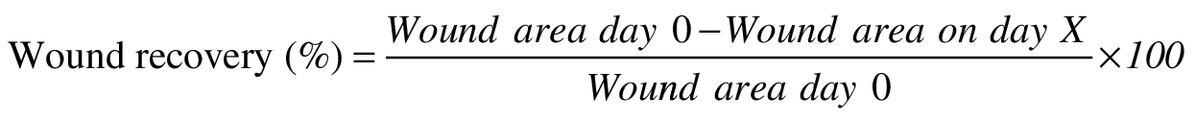
Where X represents day 4, 7, 10 or 14.
Statistical analysis
The differences in quantitative data were assessed by one-way ANOVA followed by the Dunnett’s multiple comparisons test analysis. The data are expressed as mean ± SD. The Kruskal–Wallis test was used to analyze nonparametric histopathological scores and differences among groups are reported as median. p-values < 0.05 were considered significant.
Results
Characteristics of lecithin liposomes
The average size (radius) of egg-ls and soy-ls was 115 ± 6 and 110 ± 4 nm, respectively, and both of them had a low polydispersity index (0 > 0.4) that estimated using dynamic light scattering analysis, as shown in Figure 1. Also, microscopically evaluation of egg-l and soy-l suspensions confirmed the formation of spherical and oval-shaped vesicles under high power field observation.

Figure 1. The representative dynamic light scattering analysis of lecithin liposome suspensions.
(A) Soy-l. (B) Egg-l.
Scavenging activity of the egg-l & soy-l
The results showed that increased concentrations of both lecithin liposomes (from 1 to 10 mg/ml) exhibited DPPH radical-scavenging activity in the range of 28.75–84.9% (Figure 2). As shown in Figure 2, the DPPH-scavenging activity of different concentrations of egg-l increased significantly (p < 0.0001) compared with equal concentration ranges of soy-l. Therefore, it is concluded that radical scavenging activity by lecithins has a concentration dependency and amplified with increasing lecithin contents.

Figure 2. ABTS and DPPH radical-scavenging capacities at the different concentrations of liposomal lecithins.
#Statistically significant differences p < 0.0001.
According to the results in Figure 2, there was a strong association between increasing the concentration of liposomal lecithins and ABTS radical scavenging activity. The ABTS scavenging activity of egg-l at 5 mg/ml (74.14 ± 0.84%; p < 0.01) and 10 mg/ml (84.9 ± 1.6%; p < 0.0001) was significantly higher than the scavenging activity of equal concentrations of soy-l (Figure 2).
Effective dose of lecithin liposomes
In our primary study, effective dose of both lecithin liposomes was estimated on six groups of three animals on wound area reduction (Figure 3). Although, both soy-l and egg-l exhibited an increasing trend in concentrations of 5 and 10% in wound healing, but there was not found any significant effect compared with control group. While topical application of egg-l 15% showed significantly effective (p < 0.05) in wound-healing percentage compared with control group during 10 days experiment. Concerning Figure 3, egg-l showed also effective on wound recovery rather than relative doses of soy-l.

Figure 3. Estimation of the effective dose of the different concentrations of soy-l and egg-l with n = 3 and mean ± standard deviation in wound-healing percentage.
Topical application of egg-l 15% was significantly effective compared with control during the 10-day experiment.
*p < 0.05.
Histological outcomes by egg-l & soy-l treatment
According to Figure 4, the number of neutrophils in both egg and soy lecithin groups and the phenytoin group was significantly (p < 0.0001) lower than the control group at all time points. In our study, topical administration of liposomal lecithins increased macrophages markedly on days 4 and 7, while it decreased the number of macrophages significantly compared with control wound sections on the 14th day (p < 0.0001) (Figure 4).

Figure 4. Histopathology diagram of wounds biopsy from the experimental groups at days 4, 7 and 14 of the study.
Significant difference values obtained from treated groups versus control were expressed by *p < 0.05; **p < 0.01; ***p < 0.001; and #p < 0.0001.
The initiation of a significant (p < 0.0001) fibroblast proliferation by treatment with lecithin liposomes on the 4th day indicated that the proliferative phase occurred earlier in treated groups while the control group was still in the inflammatory phase (Figure 4 & 5).
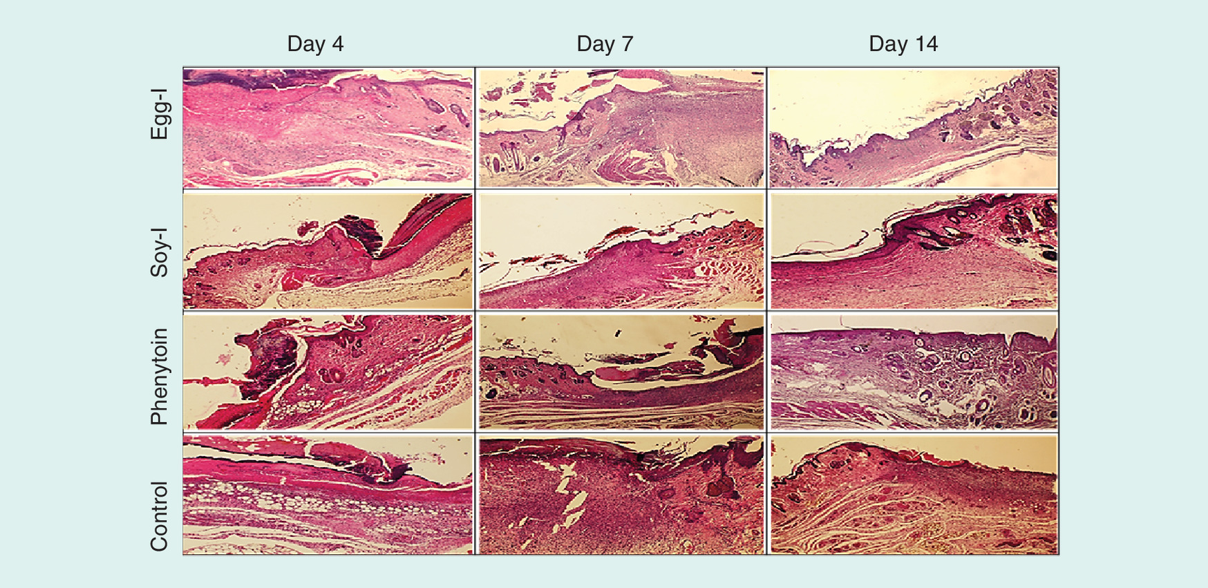
Figure 5. Photography of hemotoxylin and eosin staining related to sections of experimental groups (low power field).
Day 4, the main phenomenon of the sections is inflammation in wound bed. Day 7, granulation tissue, immature epithelialization and, collagen fibers proliferate and appear in neotissues. Day 14, complete epithelialization, considerable collagen fibers as well as granulation tissue are observed in egg-l group, which confirm reconstruction phase.
In this study, as shown in Figures 4 and 5, topical administration of lecithin liposomes once daily increased the number of fibroblasts in treated groups significantly (p < 0.0001) compared with the control group during 14 days. Phenytoin cream also had a significant effect on the proliferation of fibroblasts (p < 0.0001) similar to lecithin liposomes.
Endothelial cells, as another parameter of wound assessment, increased significant (p < 0.001) in both lecithin liposomes and phenytoin groups versus control group on days 4, 7 and 14 of the investigation (Figure 4 & 5).
According to the results by Mason’s trichrome staining (Figures 6 & 7), a significant (p < 0.001) increase of collagen deposition and re-epithelialization was in egg-l treated group in comparison with the control group on day 14 day of the experiment. Moreover, the median score of wound sections in the soy-l group showed significant (p < 0.05) re-epithelialization in the epidermis (Figure 6). However, phenytoin did not change (p > 0.05) collagen deposition and re-epithelialization indices significantly compared with the control on day 14 (Figure 6 & 7).

Figure 6. Histopathological scores of collagen deposition and re-epithelialization obtained from experimental groups at the 14th day of wounding.
The score analysis of the wound tissues (n = 6) as median ± standard deviation expressed with error bars (min–max). Significant difference values obtained from lecithins liposome versus control group were expressed by *p < 0.05; ***p < 0.001.
Figure 7. Masson trichrome staining of skin sections associated with rats subjected to open field wound during 14-day treatment.
(A) Representative section of a normal skin. (B) Photography of wound section of egg-l group demonstrates a complete epithelialization (red arrow), abundant collagen fibers (white arrow), and deposition of fibroblast cells (blue arrow) superior phenytoin and untreated groups. (C) Phenytoin-treated group. (D) Untreated group (control), Masson trichrome, (10×).
Wound area reduction by liposomal lecithins
As shown in Figure 8A, wound surface decreased significantly following treatment with egg-l (96% ± 1.2; p < 0.0001) and soy-l (94% ± 1.5; p < 0.0001) in compared with control (82% ± 5.8) after 14 days. Early wound contraction was also observed significantly (p < 0.01) with egg and soy lecithin liposomes 10 days after injury in rats (Figure 8B). Furthermore, phenytoin showed only a significant (p < 0.05) wound recovery percentage compared with control group at day 14 of the experiment. Treatment with phenytoin drug exhibited a delay effect on wound area reduction with number of 88% ± 2.1 in comparison with both lecithinic liposomes in this experimnt.

Figure 8. Representative wound recovery outcomes associated with individual drugs.
(A) Photograph of the wound appearances corresponding to the treated groups at relative days. (B) The percentage of wound recovery was observed statistically significant (as mean ± standard deviation, n = 6) compared with control group during 14-day treatment of rats with phenytoin, egg-l 15% and soy-l 15%. *p < 0.05; **p < 0.01; ****p < 0.001.
Discussion
DPPH, as a stable free radical, has an unpaired valence electron that is susceptible to reaction through transferring an electron to the donor molecule [18]. Therefore, unsaturated hydrocarbons of fatty acid chains and phosphate groups of phospholipids in lecithin may have a tendency to share their electrons or hydrogen to react with the free radical DPPH [19]. In addition, we used the ABTS test to determine the free radical scavenging activity of lecithin liposomes, which is used to estimate hydrogen-donating compounds and/or chain-breaking antioxidants [19,20].
The study of Wang has been shown that egg-l had more reactivity in oxidative state than soy-l under the experimental conditions [21]. It is proposed that lipid oxidation is a process that usually initializes from unsaturated fatty acids. Regarding fatty acid composition, egg phospholipids contain only 16.2% linoleate but more long-chain polyunsaturated fatty acids than soy phospholipids that comprise 56.6% linoleate, resulting in egg-l has higher oxidation susceptibility than soy-l [21].
Types of lecithin may have different oxidative stability not only because of their difference in fatty acid composition but also because of their phospholipid class composition and their charge. According different reports, egg-l had more negative Ζ potential than soy lecithin that may cause lipid oxidation of egg-l rather than soy-l in oxidative experiments [21,22].
Lecithins, alone or in combination with bioactive antioxidants, have been used to inhibit the impact of free radicals on biomaterial destruction [3,15,23]. Therefore, it can be concluded that the scavenging activity of lecithin liposomes might affect the improvement of wound through inhibiting the induction of oxidative stress due to free radicals.
In our study, egg-l treatment was associated with better wound-healing outcomes compared with soy-l and even phenytoin cream, which may be related to its greater antioxidant capacity. The results of a previous study by Sturm and Dignass in 2002 showed that phosphatidylcholine extracted from egg yolk possesses anti-inflammatory and antioxidant effects and prevents oxidative stress in gastrointestinal wound repair [8]. Furthermore, it has been confirmed that increase of the antioxidant capacity in a chronic wound can accelerate the wound-healing process [24,25]. Although some levels of ROS can be detected in the wound fluid [19,26] and enhance defence against pathogens in the early stages of inflammation [26,27], excessive amounts of ROS are toxic in chronic or nonhealing wounds because of their high reactivity with cell components such as the phospholipid, DNA and proteins [11].
The wound-healing process usually has three phases, including inflammation, proliferation and remodeling (reconstruction) [10,16]. It is recommended that prolonged inflammation should be inhibited in chronic or nonhealing ulcers to prevent possible injury to the organs or cancer and neoplasia formation [28].
Our finding showed that lecithin liposomes might play a modulatory role in inflammation by stimulation of the migration macrophages and inhibition of the production of inflammatory cytokine at early stages of the wound [8,9]. It has been confirmed that liposomes can be recognized as a target for macrophage and dendritic cells for phagocytosis [29] while their phospholipids could inhibit the production of proinflammatory cytokines [8,9,30]. In the second phase of wound healing, new tissue formation starts while the levels of immune cells and pro-inflammatory cytokines decrease in the wound area [11].
Moreover, our important finding in wound treatment by liposomal lecithin was angiogenesis, which plays a key role in the reparative dermis to support the capillary growth, collagen development and granulation tissue formation at the injury site [10,16].
According to our results from Mason’s trichrome staining, lecithin liposomes as comparable with phenytoin drug could potentially enhance fibroblast cells formation, collagen deposition and endothelial cell growth, which might be associated with their antioxidative capacity. As described in previous reports, the use of low molecular weight antioxidants enhances cellular proliferation and collagen synthesis at the wound site and reduces the levels of lipid peroxides [24,27,31]. In this study, the effect of lecithin liposomes on wound re-epithelialization suggested that differentiation of epithelial cells could possibly develop at an ideal rate, resulting in the formation of a normally thick epidermis in the remodeling stage [16].
We used phenytoin cream as the positive control because the mechanism of phenytoin drug showed the acceleration of wound healing. It has been investigated in pressure sores, diabetic ulcers, burns and etc., in clinical and animal studies. Phenytoin-treated open wounds have shown neovascularization, collagenization, granulation tissue enhancement and decreased polymorphonuclear cell infiltration as well as wound area reduction [16,32]. Moreover, in our experiment, treatment with lecithin liposomes exhibited a considerable wound area reduction, rather than with phenytoin cream, which could be due to acceleration of collagen deposition into the lesion site [10].
Regarding the outcomes of lecithin liposomes that were majorly better than phenytoin drug on wound healing, there is a suggestion that liposomal lecithin might be applicable in treatment of wound injuries in clinical study.
Conclusion
Our findings indicated that liposomal soy and egg lecithin may potentially improve wound-healing through scavenging activities. Local administration of liposomal lecithin 15% had a better effect than phenytoin drug on different aspects of wound repair at the early stage.
•
Liposomal egg lecithin has a scavenging activities rather than soy lecithin liposomes.
•
Liposomal lecithin may potentially improve wound-healing trough scavenging free radicals.
•
Topical administration of egg lecithin liposomes can improve skin wound more potent than soy lecithin liposomes in rats.
•
Lecithin liposomes might play a modulatory role in inflammation at the early stage of wound.
•
Local administration of lecithin liposomes could potentially enhance fibroblast cells formation, collagen deposition and endothelial cell growth in rats.
•
Lecithin liposomes indicated that differentiation of epithelial cells could possibly develop at an ideal rate, resulting in the formation of a normal thick epidermis.
•
Wound area reduction, another important index in wound repair, was observed after 14-day topical treatment with lecithin liposomes.
•
Liposomal lecithin 15% acted better than phenytoin cream in wound repair.
Financial & competing interests disclosure
This research has been supported by Tehran University of Medical Sciences and Health Services grant (grant number 94-03-158-30179). The authors have no other relevant affiliations or financial involvement with any organization or entity with a financial interest in or financial conflict with the subject matter or materials discussed in the manuscript apart from those disclosed.
No writing assistance was utilized in the production of this manuscript.
Ethical conduct of research
The authors state that they have obtained appropriate institutional review board approval or have followed the principles outlined in the Declaration of Helsinki for all human or animal experimental investigations.
References
Papers of special note have been highlighted as: • of interest
1.
Lagace TA, Ridgway ND. The role of phospholipids in the biological activity and structure of the endoplasmic reticulum. Biochim. Biophys. Acta 1833(11), 2499–2510 (2013).
2.
Van Hoogevest P. Review – an update on the use of oral phospholipid excipients. Eur. J. Pharm. Sci. 108, 1–12 (2017).
3.
Pan Y, Tikekar RV, Nitin N. Effect of antioxidant properties of lecithin emulsifier on oxidative stability of encapsulated bioactive compounds. Int. J. Pharm. 450(1-2), 129–137 (2013).
• Antioxidant properties of lecithin on oxidative state as an antioxidant.
4.
Bulbake U, Doppalapudi S, Kommineni N, Khan W. Liposomal formulations in clinical use: an updated review. Pharmaceutics 9(2), (2017) (Epub ahead of print).
5.
More MI, Freitas U, Rutenberg D. Positive effects of soy lecithin-derived phosphatidylserine plus phosphatidic acid on memory, cognition, daily functioning, and mood in elderly patients with Alzheimer’s disease and dementia. Adv. Ther. 31(12), 1247–1262 (2014).
• Positive effects of soy lecithin-derived phospholipids on neurological impairment.
6.
Wat E, Tandy S, Kapera E et al. Dietary phospholipid-rich dairy milk extract reduces hepatomegaly, hepatic steatosis and hyperlipidemia in mice fed a high-fat diet. Atherosclerosis 205(1), 144–150 (2009).
7.
Na J-Y, Song K, Kim S, Kwon J. Hepatoprotective effect of phosphatidylcholine against carbon tetrachloride liver damage in mice. Biochem. Biophys. Res. Commun. 460(2), 308–313 (2015).
• Hepatoprotective effect of phosphatidylcholine through inhibition of oxidative stress.
8.
Sturm A, Dignass AU. Modulation of gastrointestinal wound repair and inflammation by phospholipids. Biochim. Biophys. Acta 1582(1-3), 282–288 (2002).
9.
Miranda DT, Batista VG, Grando FC et al. Soy lecithin supplementation alters macrophage phagocytosis and lymphocyte response to concanavalin A: a study in alloxan-induced diabetic rats. Cell. Biochem. Funct. 26(8), 859–865 (2008).
• Properties of soy lecithin supplementation on immunomodulation of wound induced diabetic and wound repair.
10.
Guo S, Dipietro LA. Factors affecting wound healing. J. Dent. Res. 89(3), 219–229 (2010).
11.
Schafer M, Werner S. Oxidative stress in normal and impaired wound repair. Pharmacol. Res. 58(2), 165–171 (2008).
• Importance inhibition of oxidative stress in treatment and wound repair.
12.
Roy P, Amdekar S, Kumar A, Singh R, Sharma P, Singh V. In vivo antioxidative property, antimicrobial and wound healing activity of flower extracts of Pyrostegia venusta (Ker Gawl) Miers. J. Ethnopharmacol. 140(1), 186–192 (2012).
• Antioxidative property, antimicrobial and wound-healing activity of natural extracts.
13.
Kianvash N, Bahador A, Pourhajibagher M et al. Evaluation of propylene glycol nanoliposomes containing curcumin on burn wound model in rat: biocompatibility, wound healing, and anti-bacterial effects. Drug. Deliv. Transl. Res. 7(5), 654–663 (2017).
14.
Taheri A, Sabeena Farvin KH, Jacobsen C, Baron CP. Antioxidant activities and functional properties of protein and peptide fractions isolated from salted herring brine. Food Chem. 142, 318–326 (2014).
15.
Hosseini SF, Ramezanzade L, Nikkhah M. Nano-liposomal entrapment of bioactive peptidic fraction from fish gelatin hydrolysate. Int. J. Biol. Macromol. 105(Pt 2), 1455–1463 (2017).
16.
Takzaree N, Hadjiakhondi A, Hassanzadeh G, Rouini MR, Manayi A, Zolbin MM. Transforming growth factor-beta (TGF-beta) activation in cutaneous wounds after topical application of aloe vera gel. Can. J. Physiol. Pharmacol. 94(12), 1285–1290 (2016).
17.
Abramov Y, Golden B, Sullivan M et al. Histologic characterization of vaginal vs abdominal surgical wound healing in a rabbit model. Wound Repair Regen. 15(1), 80–86 (2007).
18.
Sharma OP, Bhat TK. DPPH antioxidant assay revisited. Food Chem. 113(4), 1202–1205 (2009).
19.
Alemán A, Giménez B, Montero P, Gómez-Guillén M. Antioxidant activity of several marine skin gelatins. LWT-Food Sci.Techno. 44(2), 407–413 (2011).
20.
Ojha N, Roy S, He G et al. Assessment of wound-site redox environment and the significance of Rac2 in cutaneous healing. Free Radic. Biol. Med. 44(4), 682–691 (2008).
21.
Wang G, Wang T. Oxidative stability of egg and soy lecithin as affected by transition metal ions and pH in emulsion. J. Agric. Chem. 56(23), 11424–11431 (2008).
• Effect of chemical composition of lecithin of soy and egg on their antioxidant capacity and scavenging activity.
22.
Sørensen A-DM, Haahr A-M, Becker EM et al. Interactions between iron, phenolic compounds, emulsifiers, and pH in omega-3-enriched oil-in-water emulsions. J. Agric. Chem. 56(5), 1740–1750 (2008).
23.
Judde A, Villeneuve P, Rossignol-Castera A, Le Guillou A. Antioxidant effect of soy lecithins on vegetable oil stability and their synergism with tocopherols. J. Am. Oil Chem. Soc. 80(12), 1209–1215 (2003).
24.
Panchatcharam M, Miriyala S, Gayathri VS, Suguna L. Curcumin improves wound healing by modulating collagen and decreasing reactive oxygen species. Mol. Cell. Biochem. 290(1-2), 87–96 (2006).
25.
Pazyar N, Yaghoobi R, Rafiee E, Mehrabian A, Feily A. Skin wound healing and phytomedicine: a review. Skin Pharmacol. Physiol. 27(6), 303–310 (2014).
26.
Roy S, Khanna S, Nallu K, Hunt TK, Sen CK. Dermal wound healing is subject to redox control. Mol. Ther. 13(1), 211–220 (2006).
27.
Musalmah M, Nizrana MY, Fairuz AH et al. Comparative effects of palm vitamin E and alpha-tocopherol on healing and wound tissue antioxidant enzyme levels in diabetic rats. Lipids 40(6), 575–580 (2005).
28.
Cerutti PA, Trump BF. Inflammation and oxidative stress in carcinogenesis. Cancer Cells 3(1), 1–7 (1991).
29.
Kelly C, Jefferies C, Cryan SA. Targeted liposomal drug delivery to monocytes and macrophages. J. Drug. Deliv. 2011, 1–11 (2011).
30.
Hashioka S, Han YH, Fujii S et al. Phosphatidylserine and phosphatidylcholine-containing liposomes inhibit amyloid beta and interferon-gamma-induced microglial activation. Free Radic. Biol. Med. 42(7), 945–954 (2007).
31.
Senel O, Cetinkale O, Ozbay G, Ahcioglu F, Bulan R. Oxygen free radicals impair wound healing in ischemic rat skin. Ann. Plast. Surg. 39(5), 516–523 (1997).
32.
Bhatia A, Prakash S. Topical phenytoin for wound healing. Dermatol. Online J. 10(1), 5 (2004).
• Situation and benefits of natural product on wound healing.
Information & Authors
Information
Published In
Pages: 633 - 643
PubMed: 31116027
Copyright
© 2019 Future Medicine Ltd.
History
Received: 18 November 2018
Accepted: 18 February 2019
Published online: 22 May 2019
Keywords:
Topics
Authors
Metrics & Citations
Metrics
Article Usage
Article usage data only available from February 2023. Historical article usage data, showing the number of article downloads, is available upon request.
Citations
How to Cite
In vitro antioxidant activity and in vivo wound-healing effect of lecithin liposomes: a comparative study
. (2019) Journal of Comparative Effectiveness Research. DOI: 10.2217/cer-2018-0128
Export citation
Select the citation format you wish to export for this article or chapter.
Citing Literature
- Bhagavathi Sundaram Sivamaruthi, Natarajan Suganthy, Periyanaina Kesika, Khontaros Chaiyasut, Rungaroon Waditee-Sirisattha, Wandee Rungseevijitprapa, Chaiyavat Chaiyasut, Phytochemical-Loaded Nanotherapeutics in Cosmetic Surgery Wound Healing: A Narrative Review, Cosmetics, 10.3390/cosmetics13030111, 13, 3, (111), (2026).
- Lolyta Fitri Mustati, Krishnan Raguvaran, Harris Antonius, Tia Okselni, Sofna DS. Banjarnahor, Yuli Widiyastuti, Yuandani Yuandani, Bayu Eko Prasetyo, Eldiza Puji Rahmi, Marissa Angelina, Rizna Triana Dewi, Nurul Arfiyanti Yusuf, Abdi Wira Septama, Nano-transfersomal hydrogel of Curcuma xanthorrhiza essential oil: Formulation, characterization, and evaluation of their bioactivities in vitro and in vivo, Journal of Drug Delivery Science and Technology, 10.1016/j.jddst.2026.108090, 118, (108090), (2026).
- Dyandevi Mathure, Vaishnavi Bhise, Malati Salunke, Gaurav Borse, Hemantkumar Ranpise, Rajendra Awasthi, Curcumin–piperine liposomal hydrogel formulation for topical management of atopic dermatitis and associated skin inflammation: optimization, in vitro and in vivo studies, Inflammopharmacology, 10.1007/s10787-026-02144-2, 34, 3, (1851-1870), (2026).
- Tauseef Ahmad, Tasmina Kanwal, Khadija Rehman, Ali Asgher Shuja, Abdul Jabbar, Wajeeha Muzafar, Shafi Ullah, Farhat Ullah, Shabana Usman Simjee, Muhammad Raza Shah, Design and Development of a New Lecithin Containing Alkylated Biotin Functionalized Naringin Loaded Self-Nano-Emulsifying Drug Delivery System for Enhanced Anticancer and Antioxidant Activities, BioNanoScience, 10.1007/s12668-025-02397-1, 16, 2, (2026).
- Darshan Ramesh, Sindhu Abraham, Megha Krishnappa, Bharath Srinivasan, Sterile thermoresponsive formulations for emergency management of burns, Materials Today: Proceedings, 10.1016/j.matpr.2022.09.036, 117, (1-9), (2026).
- Zunaira Afzal, Sadia Rafique, Anam Hanif, Therapeutic targets of wound healing, Polyphenols in Wound Healing, 10.1016/B978-0-443-45193-5.00011-9, (61-78), (2026).
- Aniseh Mardanpoor Moghadam, Sonia Fathi-karkan, Fatemeh Tanhaye Kalate Sabz, Sahar Shariatnia, Nanostructured lipid carriers for l-carnitine delivery: physicochemical characterization and effects on asthenozoospermic sperm function in vitro, Scientific Reports, 10.1038/s41598-025-33229-7, 16, 1, (2025).
- Aldrey Nathália Ribeiro Corrêa, Naiara Jacinta Clerici, Aline Aniele Vencato, Adriano Brandelli, Nano-coatings with Baccharis dracunculifolia essential oil and nerolidol as an antifungal and antioxidant strategy in post-harvest strawberries, Biocatalysis and Agricultural Biotechnology, 10.1016/j.bcab.2025.103830, 70, (103830), (2025).
- Aleksandar Marinković, Đura Nakarada, Miloš Marinković, Hadi Waisi, Vladislav Živanić, Arcadio Vazquez, Miloš Mojović, From Analytical Profiling to Liposomal Delivery: Cannabinol as a Model for Antioxidant Encapsulation and Diffusion Enhancement, Molecules, 10.3390/molecules30163433, 30, 16, (3433), (2025).
- Huang-Kai Guo, Gan-Hao Yang, Lin-Wen Wu, Jia-Qi Hong, Wei Huang, Ka-Hing Wong, Lei Chen, Qiong-Qiong Yang, Bo-Bo Zhang, Effects of ethanol extracts of Lycium chinense berry fruit on the production, structure and bioactivity of exopolysaccharides in Cordyceps militaris, International Journal of Biological Macromolecules, 10.1016/j.ijbiomac.2025.145409, 319, (145409), (2025).
- See more